


The University of Padua
The University of Padua was founded around 1220 by students and professors who left the University of Bologna to seek greater academic freedom (“Libertas scholastica”) after a brief attempt at establishment in Vicenza. It is the second-oldest university in Italy, and the fifth-oldest university still active in the world.
In 1399 the institution was divided in a Universitas Iuristarum for civil law and for Canon law, and Universitas Artistarum for astronomy, dialectic, philosophy, grammar rhetoric and medicine.
Teaching medicine began in 1222. At the time it was the most renowned school for its long-standing tradition in medicine and anatomy, linked to great figures such as Vesalius, the “father of modern anatomy,” Padua's famous anatomical theatre, the oldest surviving example of its kind in Europe, has attracted artists and scientists to study the human body at public dissections since it was built by Girolamo Fabrici d’Acquapendente in 1595. Its golden age was characterised by the introduction of empirical and experimental methods into theory-based teaching.
Andreas Vesalius
Andreas Vesalius was born Andries van Wesel in 1514 in Brussels. His great-grandfather, Jan van Wesel, received a medical degree from the University of Pavia and taught medicine at the University of Leuven, and his grandfather was the Royal Physician to Emperor Maximilian.
In 1533, Vesalius entered the University of Paris where he studied Galen’s theories and developed an interest in anatomy, examining excavated bones in the Cemetery of the Innocents and assembling his first skeleton.
Forced to come back to Leuven, he graduated in medicine there in 1537. On the same day, he was offered the chair of surgery and anatomy (explicator chirurgiae) at the University of Padua. He would also become a guest lecturer at the Universities of Bologna and Pisa. While in Venice during this period, he met the illustrator Johan van Calcar, with whom he would produce unprecedentedly detailed anatomical illustrations.
Anatomy had traditionally been taught via classical texts and animal dissection performed by a surgeon under the lecturer’s verbal direction, and Galen's statements were considered indisputable. Vesalius, on the contrary, taught through direct observation and hands-on participation, performing dissections himself and encouraging his students to do the same. He created, in collaboration with van Calcar, six large woodcut posters for the classroom, which, upon learning that some of them had been widely copied by other teachers, he published under the title Tabulae anatomicae sex.
In contrast to Galen, who worked essentially on animals, Vesalius was able, thanks to a judge at the Padua criminal court who took an interest in his work, to procure human cadavers – executed criminals – for dissection.
In 1543 Vesalius published Andrea Vesalii suorum de humani corporis fabrica librorum epitome, known as the De Humani Corporis Fabrica, an unprecedented work in medical history that established anatomy as a modern, descriptive science. In these seven books Vesalius presents a careful examination of the organs as well as the complete structure of the human body, which would have been impossible without Renaissanace-era advances in visual representation and precise woodcut printing techniques which allowed for the production of superior quality illustrations.
The Fabrica emphasises dissection and the “anatomical” view of the body, showing its internal function and structure, with the organs arranged in three-dimensional space. It contains pictures of organs on two pages, allowing for the creation of three-dimensional diagrams by cutting out the images of organs and pasting them on the duplicated figures.
The seven books of the Fabrica are organised as follows: (1) the Bones and Cartilages, (2) the Ligaments and Muscles, (3) the Veins and Arteries, (4) the Nerves, (5) the Organs of Nutrition and Generation, (6) the Heart and Associated Organs, and (7) the Brain. In the book about veins and arteries there are, among other observations, descriptions of the azygos veins and the canal called the ductus venosus in the foetus between the umbilical vein and the vena cava. In the book about the heart, he describes the strong attachment of the pericardium to the diaphragm, the shape and orientation of the ventricles of the heart, and the semilunar valves. He closes the book with a chapter on the method and technique of heart dissection.
The book sparked public interest in dissections and led to the establishment of anatomical theatres in many other European cities. Over 250 illustrations, generally attributed to students from “the studio of Titian”, show great artistic merit and accuracy, and were far superior to the illustrations in publication at the time, which had not been made by actual anatomy professors. These woodcut blocks were transported to Basel and published by one of the leading printers of the time, Johannes Oporinus, who included Vesalius' notes for the illustrators due to the interest he found they added to the work.
Some of Galen's inaccuracies, i.e., the notion that the great blood vessels originate in the liver, and that the heart contains a porous cardiac interventricular septum, were disproven in the Fabrica. Vesalius deemed Galen's observations erroneous because they were based on animal, not human, dissection. About 300 of Galen’s errors were corrected by Vesalius thanks to his observation of human bodies. Even so, some galenic misunderstandings were perpetuated by Vesalius, such as the idea that veins and arteries carried different types of blood, which was widely believed in Europe until proven false by William Harvey in 1628. Nonetheless, Vesalius' work on the vascular and circulatory system was an outstanding contribution to modern medicine, opening new methods and new perspectives.
Matteo Realdo Colombo
Matteo Realdo Colombo (1515–1559) trained as a surgeon for several years under the Venetian Giovanni Antonio Lonigo. In 1538, he arrived in Padua, where he studied medicine and anatomy. He became a close friend of Vesalius, perhaps assisting him at a dissection, and teaching in his place when the great anatomist travelled to Basel. Vesalius attributed many of the discoveries in De Fabrica to Colombo, whom he referred to as a "very good friend”.
Colombo rejected the work of previous anatomists that had been presented without proof, with particular attention to Galen. Until then, anatomists such as Galen and Vesalius had examined blood vessels separately from the organs. In contrast, Colombo analysed the vessels together with the organs they are connected to and was able to hypothesise the flow of blood to and from each organ, supporting his discovery of the pulmonary circulation of the blood.
Moreover, he considered the lungs to be separate from the heart, assigning them a role defined by respiration. This approach reinforced his criticisms of Galen. He affirmed that the ventricular septum is not permeable, rejecting the possibility of the direct passage of blood from the right ventricle to the left. Colombo’s discovery of the pulmonary circulation, which paved the way for William Harvey’s discovery of circulation years later, was due to finding only blood – and no air – in the pulmonary veins of dogs and other animals during vivisection. In his model, venous blood circulated from the heart to the lungs thorough the pulmonary artery, where it was mixed with air and then returned to the heart via the pulmonary veins. The discovery that the principal activity of the heart is contraction, rather than dilation, as previously thought, allowed for this new vision of circulation.
Colombo's anatomical work is grouped into 15 different books, each of which covers information concerning a different part of the body. Notably, the lungs are not included in the book about the heart but are considered separately.
Giovanni Battista Morgagni
Giovanni Battista Morgagni (1682–1771) studied philosophy and medicine at the University of Bologna and graduated from both faculties in 1701. When Valsalva went to Parma University, Morgagni succeeded him in anatomical demonstrations and earned a lofty reputation in Bologna, having published the communications to the academy in 1706 under the title of Adversaria anatomica.
He transferred to Padua in 1712, teaching medicine there with much success until his death, and being promoted to the chair of anatomy. During his first years at Padua, Morgagni published five more volumes of the Adversaria anatomica.
In 1761, at the age of eighty, he released the great work which would make pathological anatomy a science, using it to look for the origins and causes of diseases and opening new channels of precision into medicine: the De Sedibus et causis morborum per anatomen indagates. This work comprehends five books addressing the morbid conditions of the body a capite ad calcem and contains the records of more than 600 dissections.
Figure 1. Morgagni - Fifth book of the ‘De Sedibus’.
(Public domain, via Wikimedia Commons)
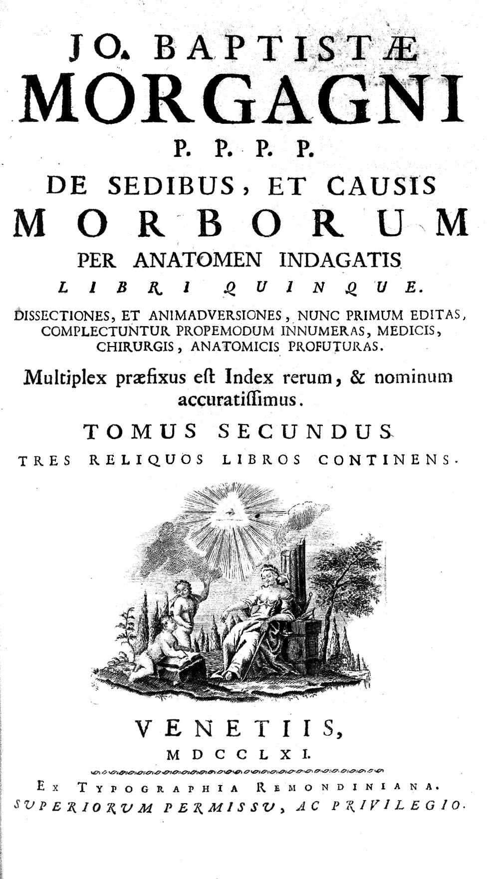
Some of these are written with a precision of statement and exhaustiveness of detail that has gone unsurpassed until now. The various symptoms of a malady and other antecedent circumstances included in the descriptions are fairly complete. Of the 70 letters grouped into the 5 volumes of the De Sedibus, 13 are devoted to diseases of the heart and vessels. In these, he describes various congenital heart diseases – cases of aortic coarctation in two letters, cases of atrial septal defects in others, as well as others that discuss the Trilogy of Fallot and a patent ductus Botalli.
Elsewhere, in great detail, he depicts patients who died of valve diseases and their consequences on the ventricles and atria, managing to show a case of functional tricuspid regurgitation due to right ventricle dilation. He analyses patients with coronary artery diseases, identifying coronary calcification, cardiomyopathy due to myocardial infarction, and a left ventricle rupture due to a myocardial infarction in a case of sudden death. Arrhythmias also are treated in the De Sedibus, with cases of bigeminy and of bradycardia (atrioventricular block or Tachy-Brady Syndrome?) in a patient with recurrent syncope with a heart rate of 22 bpm followed by tachycardia. This was later named “Morgagni-Adams-Stokes syndrome”.
Additionally, he included many patients with pericardial diseases, such as pericardial effusions, haemopericardium, pericardial tumours, aortic aneurysms (mainly luetic), aortic dissections, and aortic atheroma. Finally, he was able to develop an integrated view of symptoms like dyspnoea, nocturnal paroxysmal dyspnoea, ankle oedema, oliguria, arrhythmias, hepatomegaly, pleural effusion and ascites with dilated sick hearts, showing a clear picture of heart failure.
Lazzaro Spallanzani
Lazzaro Spallanzani (1729–1799) studied law at Bologna University, became professor of logic, mathematics, and metaphysics at the University of Reggio in 1754, and in 1768, accepted an invitation to the chair of natural history at the University of Pavia.
In 1785 he was invited to the University of Padua. Here, thanks to the recent invention of the microscope, he was able to demonstrate the microscopic circulation into the capillary network in warm-blooded animals. He also studied the slowing of the blood during pathology processes, the effects of gravity on the vascular system, changes in blood vessel diameter, and blood flow velocity, demonstrating that it is quickest during systole.
Antonio Scarpa
Antonio Scarpa (1752–1832) became a Doctor of Medicine in 1770 in Padua and was a student of Morgagni. in 1783 he became professor of anatomy at the University of Pavia. In 1804, he wrote his main work, Sull’aneurisma (On the aneurysm), which gives a detailed description of the slow degeneration of the intima of the vessel, leading to its progressive deterioration, dilation, and finally rupture.
After the fall of the Serenissima Republic of Venice in 1797, Padua endured the domination of the French and then the Austrians, and experienced Italy’s tumultuous Risorgimento period. These events led to the beginning of a dark age that did not spare the University, which did recover its standing until the late 19th and early 20th centuries with the addition of new faculties.
William Harvey
It should be remembered that it was from Padua University that William Harvey graduated in 1602, who would publish in 1628 his Exercitatio Anatomica de Motu Cordis et Sanguinis in Animalibus, commonly known as De Motu Cordis, basing his conclusions on the influence of Michael Servetus and, above all, Andrea Cesalpino. Note that an upcoming volume of Cardiopractice will be specifically dedicated to William Harvey.
Other schools
In addition to those already mentioned, the universities of Pisa (founded in 1343), Pavia (with evidence of teaching, including by some of the anatomists discussed above, dating back to 1361), Naples (established in 1224), and Rome Sapienza (founded in 1303), were also important places of learning in Italy.
Other essential figures
As discussion of historical medical schools in Italy comes to a close, there are three more essential figures who must be included, who either passed from one university to another or worked outside of academia.
Andrea Cesalpino
Andrea Cesalpino (1524/1525–1603) studied medicine at the University of Pisa under Realdo Colombo, then travelled through Italy, went to Padua and came back to Pisa. His physiological research about blood circulation, based prevalently on philosophical concepts and not founded on anatomical research, probably led to a first hypothesis about arterial peripheral circulation. He wrote: “Sanguis fugit ad cor tanquam ad suum principium, non ad hepar aut cerebrums”, (“Blood flows to the heart as though to its source, not to the liver or the brain”), rejecting one of the most important postulates of Galen. In his 1571 work Quæstionum peripateticarum, he makes the first use of the term “circulation”, and states that blood continuously flows from the arteries to the veins by means of anastomoses called vasa in capillamenta resoluta, and that veins bring the blood to the heart which distributes it by means of the arteries “in universum corpus.” In Quæstionum medicarum, 1593, he was able to demonstrate via experimentation that venous blood flows from the periphery to the centre of the body. It may be said that he penetrated more deeply into the secrets of the circulation of the blood than any other physiologist before William Harvey.
Giovanni Maria Lancisi
Giovanni Maria Lancisi (1654–1720) graduated from the school of medicine at the University of Rome in 1672 and held the chair of anatomy at La Sapienza University until 1697. He published extensively in the field of cardiology, giving descriptions of vegetations on heart valves, of cardiac syphilis aneurysms, and also developed a classification of heart diseases. His work, De subitaneus mortibus, was published in 1707, and De motu cordis et aneurysmatibus was published posthumously in 1728. In the former, he hypothesised the causes of sudden death in the dysfunction of the brain, heart, and lungs. He distinguished merely apparent from real death by placing a candle near the nose and mouth and a container of liquid on the sternal region of the patient in order to observe respiratory function and cessation of pulsation. Nerve stimulation could also be used in the diagnosis. He also described pulmonary oedema, pulmonary embolism, and gas embolism in particular. The latter discusses subjects including the anatomy and physiology of the heart, diseases of the arteries and veins, as well as other conditions, such as right ventricle heart failure, with a precise definition of the jugular venous pulse with distention.
Leonardo da Vinci
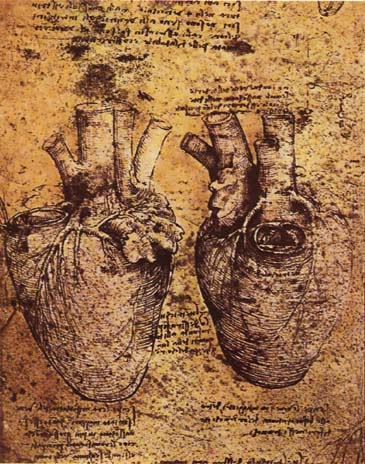
Although not linked to the academic world, it would be impossible to conclude this review of Italian cardiological history without mentioning the monumental work of Leonardo da Vinci (1452–1519). Besides his activity as a painter, draughtsman, engineer, scientist, theorist, sculptor, and architect, he was also an eminent anatomist, introducing the scientific method into anatomical observation well before Vesalius by personally conducting human dissection. He was the first to inject solidifying substances into empty organs, of which he made over 240 detailed drawings and wrote about 13,000 words towards a treatise on anatomy. He intended this work to be composed of 120 books but was unable to finish it due to the multitude of activities in the different scientific fields he was involved with. Together with the anatomical structure of the organs, he studied physiology, embryology, and comparative anatomy. He dedicated three notebooks to the heart and circulation, drawing the heart, the coronary arteries, and veins with extreme precision. He also described the ventricles and atria, found a patent foramen ovale, studied the atrio-ventricular and semilunar valves, showing the connection to the ventricles and papillary muscles of the chordae tendineae, and discovered the moderator band in the right ventricle. He carried out experiments on circulation physiology using ingenious models: for example, he constructed a glass aorta to observe the passage of blood through the aortic valve by using water and grass seeds in order to observe flow patterns. Although he understood that blood is pushed from the left ventricle in the aorta, he was ultimately unable to clarify the mechanism of the venous return in both systemic and pulmonary circulation.
Figure 2. Leonardo da Vinci – drawing of the heart.
(Public domain, via Wikimedia Commons)
Conclusion
So concludes this overview of Italian contributions to cardiovascular medicine, bringing us to the doorstep of the modern era. Though many technological advancements have been made since then and many secrets of the human body have been discovered, there still lies before us a vast expanse of knowledge to be gleaned by doctors and scientists in Italy and across the world. As we continue this pursuit of knowledge, however, we must not forget to remember how we have managed to arrive where we find ourselves now.
