In order to bring you the best possible user experience, this site uses Javascript. If you are seeing this message, it is likely that the Javascript option in your browser is disabled. For optimal viewing of this site, please ensure that Javascript is enabled for your browser.
Did you know that your browser is out of date? To get the best experience using our website we recommend that you upgrade to a newer version. Learn more.

Patent foramen ovale in patients with cryptogenic stroke: to close or not to close?

Evidence exists supporting percutaneous patent foramen ovale (PFO) closure and medical therapy versus medical therapy alone in selected patients with cryptogenic stroke (CS). However, the procedure may be associated with cardiac complications. Transoesophageal echocardiography (TOE), despite its limitations, is considered the ‘gold standard’ for PFO diagnosis but an unequivocal diagnostic algorithm for the detection of PFO and its treatment is still lacking.  The Pascal classification system recommends PFO closure plus antiplatelet therapy in selected patients aged under 60 years (essentially those with a large right-to-left shunt or an atrial septal aneurysm), without other evident cause of stroke.

Stroke

Keywords 

cryptogenic stroke; embolic stroke; patent foramen ovale; patent foramen ovale closure; stroke


Abbreviations

ASA: atrial septum aneurism

CS: cryptogenic stroke

PFO: patent foramen ovale

RA: right atrium

TCD: transcranial doppler

TOE: transoesophageal echocardiography

TTE: transthoracic echocardiography


Take-home messages

  1. PFO can in many cases be considered an anatomical variant. Patients with a PFO and without CS should be reassured that no further exams are necessary.
  2. The relationship between CS and PFO is known but remains unclear.
  3. CS diagnostic workup (AF screening – PFO Screening) - Looking for PFO in patients under 60 years of age with CS.
  4. PFO closure in CS: limitations and antithrombotic therapy.
  5. Patient selection for device closure. What to do in daily practice.


Patient-oriented messages

About one-quarter of adults have patent foramen ovale (PFO) and in the absence of symptoms this can be considered a normal anatomical variant.

Cryptogenic stroke (a stroke of undetermined origin) is a brain infarction not attributable to a definite cardioembolic event, after appropriate vascular, cardiac, and serological investigations, suspected to be a result of a cryptogenic embolism, that may be related to a PFO.

Percutaneous PFO closure with specific devices has been proposed for stroke recurrence prevention, but in some limited instances these devices may entail side effects, such as atrial fibrillation, limiting their efficacy and safety.

Careful screening to identify the patients with CS who most may benefit from PFO closure is required.


Central Figure. Relationship between PFO and CS and indications for closure.

361_Forno_FINAL FOR PUBLICATION_Central Figure.jpg


PFO as anatomical variant

The foramen ovale (FO) allows blood to shunt from the right atrium (RA) to the left atrium (LA) during foetal life. At birth, the reversal of the interatrial gradient pushes the septum primum against the septum secundum, functionally closing the FO. The anatomic closure occurs a few days after birth. However, in one quarter of adults, an incomplete apposition of the two septa creates a ‘tunnel’ between RA and LA, a patent foramen ovale (PFO).

Different anatomical variants exist. A prominent Eustachian valve may increase blood flow from the inferior vena cava (IVC) to the PFO enhancing the probability of shunting. The PFO diameter ranges in the literature from 1 to 19 mm.  The septum primum may become stretched and aneurysmal (atrial septum aneurism [ASA]), sometimes with fenestrations, increased mobility and bulging. 

PFO is detectable in a large proportion of adult subjects (10-24% on TTE, 22-39% on transoesophageal echocardiography [TOE] and 15-36% at autopsy) [1] and PFO prevalence seems to be greater in patients with cryptogenic stroke (CS). In 561 subjects with CS, an isolated PFO was found in 37%, a PFO + ASA in 46% [2]. It also seems that smaller shunts with a very large ASA entail an increased risk of stroke recurrence. In a large meta-analysis, risk factors for PFO included younger age (<50 years old), radiographic evidence of a cortical infarct and an absence of traditional vascular risk factors [3, 4].  

However, many studies following PFO patients with no history of thromboembolism have not shown an increased risk of stroke recurrence. In daily practice, PFOs are frequently found incidentally in patients without any clinical signs or symptoms. In one prospective study, a PFO was not demonstrated to be predictive of future cardiovascular events after correction for age and copathologies [5].

As a consequence, in the absence of systemic thromboembolism, a PFO should be considered an anatomical variant instead of a pathologic condition. Patients with known PFO should be reassured that neither exams nor interventions are required. Currently, there is no role for percutaneous PFO closure for primary prevention. In some instances, PFO closure could be considered deep venous thrombosis (DVT) prophylaxis and treatment, and it is advisable that scuba divers with a PFO not exceed 25-30 m of depth and use nitrox instead of compressed oxygen. For migraine prevention, the Mist Trials showed a negative result of PFO closure [6].

The relationship between cryptogenic stroke and PFO

Cryptogenic stroke (a stroke of undetermined origin), as defined by the TOAST classification [7, 8],  is a brain infarction not attributable to a clearly recognised cardioembolic event, carotid or intracranial artery disease, or other diseases after appropriate vascular, cardiac, and serological investigations. A CS is, consequently, suspected to result from a cryptogenic embolism.

Although a relationship between CS and PFO was first reported more than twenty years ago, both conditions have a high prevalence, and it is hard to prove a causal relationship. About 30% of strokes in the US and 25% in Europe are considered CS [9-11].

An Oxford meta-analysis showed a risk of stroke recurrence after CS at 1 month of 4.2% and in the National Institute of Neurological Disorders and Stroke Data Bank of 3%. The recurrence risk rose to 14-20% after 2 years, and to 33% after 5 years in Olmstead County [12,13].

Unfortunately, in recent randomised controlled trials (RCTs) on CS and PFO, the CS rate in the population and the rate of recurrent stroke following CS was lower than in previous studies, reducing their statistical power. Some studies have shown a higher rate of CS recurrence associated with ASA or atrial septum (AS) hypermobility (>10 mms) together with PFO [14, 15].

The commonly accepted pathophysiological hypothesis of the involvement of a PFO in a CS is a ‘paradoxical embolism’ of a thrombus passing through the PFO and entering the arterial circulation. Any increase in right-to-left shunting due to intermittent elevation of RA pressure (Valsalva manoeuvre, coughing, caval compression) enhances the risk. Also, thrombus formation in situ, within the PFO or on an ASA, is another potential source of thromboembolism. A large ASA can act like a net, capturing thrombi in the RA and pushing them to the PFO. Given these considerations, percutaneous PFO closure has been proposed for stroke recurrence prevention.  Multiple trials have been published with conflicting results, and areas of low evidence about PFO closure remain to be further defined.

CS diagnostic workup

Atrial fibrillation screening

Most CS, according to brain imaging (essentially cerebral magnetic resonance imaging [MRI]), are considered secondary to a cardiac thromboembolism, as undiagnosed asymptomatic atrial fibrillation (AF).

AF screening should be considered in all CS patients with a 12-lead electrocardiogram (ECG) and 24-hour Holter monitoring or other longer monitoring (48-72 hours or a 7-day ECG patch). High-risk AF patients (i.e., age >65 years, hypertension, diabetes, heart failure or cardiac structural abnormalities) require longer monitoring with an implantable loop recorder (ILR) if standard screening is negative, before a PFO closure decision can be made [16].

Echocardiographic PFO screening

Most patients with stroke or transient ischaemic stroke (TIA) have a normal transthoracic echocardiogram (TTE). When an interatrial shunt is suspected, a PFO may be shown by contrast imaging methods, with the passage of agitated saline bubbles into the left heart within 3 cardiac cycles of RA opacification.

A semi-quantification of the shunt is related to the number of bubbles shunting: small (3-10 bubbles), medium (10-20), large (>20). The contrast is generally injected via an arm vein, but sometimes blood flow within the RA is inefficient because the PFO shunt flow preferentially comes from the inferior vena cava (IVC). In cases where PFO is highly suspected, a lower extremity vein injection should be used in an alert patient capable of a Valsalva manoeuvre (Video 1 and Video 2).


Video 1. Transoesophageal contrast study, mid-oesophageal bicaval view: opacification of left atrium after injection after injection of contrast (9 cc agitated saline + 1 cc air) from superior vena cava (SVC), with evidence of severe right-left shunt (>20 bubbles).


Video 2. Transoesophageal contrast study mid-oesophageal bicaval view: almost complete opacification of left atrium after injection of contrast from SVC, during the release phase of a Valsalva manoeuvre.


The detection of the shunt may be diagnosed by contrast TTE (cTTE), contrast TOE (cTOE), or contrast transcranial Doppler (cTCD).  Sensitivity and specificity are 46% and 99% for cTTE, 89% and 92% for cTOE, and 96% and 93% for cTCD, respectively [2]. The Valsalva manoeuvre is an opportunity to increase RA pressure transiently and unmask a transient shunt across a PFO. An ESC position paper [14] algorithm recommends cTTE as first investigation to identify a PFO, and if the cTTE is normal, cTOE or cTCD can be used as alternatives.  cTCD cannot distinguish an intracardiac shunt from an intrapulmonary shunt (e.g. pulmonary arteriovenous malformations) and does not show the interatrial septum (IAS) anatomy. However, thanks to its high sensitivity, it is a reliable screening tool for detecting right-to-left shunts.

In patients with a clear contrast demonstration, cTOE can clarify the IAS anatomy, especially at the level of the fossa ovalis. An ASA is diagnosed by cTOE, finding a fixed displacement or a hypermobile fossa ovalis region of the AS toward the RA or LA, or both, exceeding 10 mm from the midline or a combined total excursion of 15 mm.

The main disadvantage of cTOE which is seen to reduce its sensitivity is the impossibility for the sedated patient to perform a Valsalva manoeuvre. A study-level meta-analysis in 2014 [17] on the accuracy of cTOE in diagnosing PFO and an interatrial shunt, compared to autopsy, cardiac surgery or catheterisation, showed a weighted sensitivity of only 89%, highlighting the lack of an available non-invasive gold standard method for the evaluation of PFO-related shunts. In selected cases it may be useful to antagonise anaesthesia.

Nakayama et al proposed an echocardiographic score to define high-risk PFO based on: large-size PFO (>2 mm in height), long-tunnel PFO (>10 mm in length), ASA, hypermobile AS, prominent Eustachian valve or Chiari’s network, large right-to-left shunt at rest and during Valsalva manoeuvre, and low-angle PFO (<10° of PFO angle from inferior vena cava). The presence of two or more high-risk PFO features (score ≥2) entails a higher risk for CS [18].

PFO closure in CS

PFO closure methods and limitations

Data from randomised trials and a meta-analysis support PFO closure in high-risk patients. These recommendations are based on the probability of the role of PFO when other aetiologies of CS have been excluded.  Many RCTs, such as the CLOSE [19], REDUCE [20], DEFENSE-PFO [21] and the RESPECT [11] with prolonged follow-up, have shown that in patients <60 years with a high-risk PFO, closure in the first 6 months after the index stroke reduces stroke recurrence but not mortality.

In several metanalysis, despite methodological limitations, consensus exists that PFO device closure should be performed in CS patients with the highest risk. No differences have been reported about superiority of a specific device.

PFO closure may have potential complications. 6.3% subjects had recurrent stroke and/or TIA, and 3.9% subjects had a residual right-to-left shunting in a cohort of 730 patients who underwent PFO closure for CS [22]. New incidence AF varies depending upon the PFO device delivered, (13% for the Cardioform, 4% for the Helex and 4% for the Amplatzer).  Moreover, rare, but devasting are device thrombosis, embolism, displacement, infective endocarditis and aortic root erosion [23]. Consequently, it is imperative that patient selection identifies those who most will benefit from the intervention.

PFO closure decision should be undertaken by a multidisciplinary team (MDT), that includes a neurologist in order to confirm that the CS is most likely due to an embolic event, that the PFO has high-risk features, to exclude causes of embolic stroke, especially AF, and to confirm anatomical eligibility for device closure and review the risk of complications. The indication should be discussed with the patient with a clear and complete explanation of the risks and benefits of the procedure, any additional medical therapy required, and any alternative options. 

Medical therapy and PFO

The alternative to PFO closure is medical therapy which should be considered in low-risk patients without indications for PFO closure, and an assessment of bleeding risk versus the PFO-stroke related relapse risk should be made. Vitamin K antagonists (VKA) may be the best choice in low bleeding risk patients but requires good compliance and adequate monitoring. Direct oral anticoagulants (DOACs) need further study to extend their use in this setting. Whilst DOACs have shown greater antiembolic protection and a reduced bleeding risk compared to VKA in patients with AF, the results compared to antiplatelet agents in CS have not been confirmed.

Antiplatelet therapy is an alternative when the risk of stroke is low. In the RESPECT trial [11], in the medical therapy arm with anticoagulant or antiplatelet therapy there was no benefit of PFO closure among the patients with indication to anticoagulation.

Patient selection for PFO closure

Patient characteristics

A young age (≤ than 60-years-old), no common cardiovascular (CV) risk factors, no other potential CV causes of thromboembolism, concomitant DVT or DVT risk factors, ASA or a large shunt association are considered as the main patient characteristics that favour a PFO closure for secondary prevention after a CS. ASA association justifies the PFO closure. There is less agreement about shunt entities without an ASA or other risk factors. However, a study meta-analysis showed a benefit in PFO closure in large shunts, also independently of ASA. The same indication is found in the CLOSE [19] and DEFENSE-PFO [21] trials in selected high-risk patients, with or without ASA.

Two risk scores have been proposed to assess the relationship between stroke and PFO. The Risk of Paradoxical Embolism (RoPE) mainly includes clinical features while the PFO-Associated Stroke Causal Likelihood (PASCAL) classification system includes both the RoPE score and the anatomical features of an ASA and large shunts (Table 1).


Table 1. RoPE Score and PASCAL Classification System.

RoPE SCORE
                  Characteristic                                                                                                                    Points
No history of:

Hypertension
Diabetes
Stroke or transient
ischaemic attack

+ 1
+ 1
+ 1

Non-smoker   +1
Cortical infarct on imaging   +1
Age (in years)

18–29
30–39
40–49
50–59
60–69
>70

+5
+4
+3
+2
+1

PASCAL classification system

High RoPE score (≥7)

High-risk PFO feature (large
shunt and/or ASA)a

Stroke related to PFO

Absent 

Absent

Unlikely

Absent

Present

Possible

Present

Absent

Possible

Present

Present

Probable

aA large shunt size is defined as >20 bubbles in the left atrium on TOE; ASA defined as >10 mm of excursion from midline. ASA: atrial septum aneurism; PASCAL: PFO-Associated Stroke Causal Likelihood; PFO : patent foramen ovale; RoPE: Risk of Paradoxical Embolism


Based on these combinations of factors, the original, extended PASCAL classification system assigns the likelihood of a causal relationship to five levels: definite, highly probable, probable, possible and unlikely. The PASCAL algorithm [24] was developed using a mixed methods approach, incorporating expert judgement, physiological and epidemiological data and the validated RoPE score.

What the guidelines say

Over the last ten years, a series of position papers and guidelines about PFO management have been published. In 2013-2016, US stroke guidelines tributed to PFO closure in CS a class 2b recommendation. After the 2017 publication of the CLOSE [19], REDUCE [20], and DEFENSE-PFO [21] trials and 3 metanalyses in 2018-2019 favouring PFO closure for CS, consensus statements from the French Society of Cardiology [14] and the European Society of Cardiology were published in 2019. Percutaneous PFO closure in CS patients with ASA or isolated PFO with large shunts was recommended [18]. The American Heart Association (AHA) stroke guidelines in 2019 underlined doubts about the quality of the RCTs and favoured PFO closure versus antiplatelet therapy, but not versus anticoagulant therapy [25].

In 2022, the Society for Cardiovascular Angiography & Interventions Guidelines recommended PFO closure in patients aged 18 to 60 years (strong recommendation, moderate certainty of evidence), suggesting that a RoPE (risk of paradoxical embolism) score ≥7 may identify patients at greater benefit [26].

Finally, in 2024 the European Stroke Organisation (ESO) Guidelines were published, developed with a strong and tight Grading of Recommendations, Assessment, Development and Evaluations (GRADE) methodology [27]. However, some questions remain unanswered and require larger evidence from RCTs as well as critical review and evaluation.

Management in daily clinical practice

Despite the aforementioned limitations, the ESO Guidelines [27] are now the reference for clinical practice in the field of CS management and PFO closure. The population, intervention, comparator, outcome (PICO) questions consider: the diagnostic performance of cTTE, cTCD and cTOE, the risk reduction of CS relapse after PFO closure plus antiplatelet agents versus antiplatelet alone, the use of the PASCAL classification in patients with different age classes, the management of anti-aggregation or anticoagulation in the short- or long-term as an alternative or in association with PFO closure.

The use of risk stratification tools was not prespecified in the PICO questions and therefore no systematic review was conducted with regard to the PASCAL classification system. Nevertheless, it was used because it was associated with a clear, demonstrated differential treatment effect, but was degraded by one level because it was not used as an inclusion criterion or stratification variable in any existing RCTs, nor was it prospectively validated.

The conclusions on PFO closure are [27]:
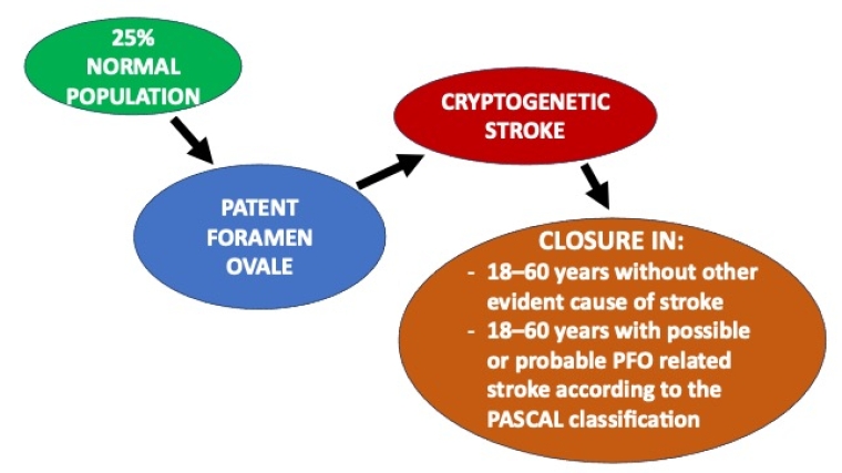
  • In patients aged 18–60 years without other evident cause of stroke but a PFO, PFO closure in selected patients is recommended in addition to antiplatelet therapy (Quality of evidence: High - Strength of recommendation: Strong for intervention).
  • In patients aged 18–60 years with possible or probable PFO related stroke according to the PASCAL classification, PFO closure in addition to antiplatelet therapy is recommended (Quality of evidence: Moderate - Strength of recommendation: Strong for intervention).
  • In patients aged 18–60 years with unlikely PFO-related stroke according to the PASCAL classification, the suggestion is against PFO closure unless there is a high probability of clinical causality enhancing the risk of paradoxical embolism (non-cerebral embolism, deep venous thrombosis and/or pulmonary embolism close to index stroke, pulmonary arterial hypertension, history of sleep apnoea or other hypoxaemic conditions associated with PFO, Valsalva at stroke onset, recent history of prolonged immobility, recent airline travel, presence of venous thrombophilia, decompressive illness in divers, platypnea-orthodeoxia syndrome or a Eustachian valve or other anatomical features on echocardiography) (Quality of evidence: Low - Strength of recommendation: Weak against intervention).
  • In patients older than 60 and younger than 18 years, no evidence-based recommendation can be provided (Quality of evidence: Very Low + Strength of recommendation: N/A).
  • Given the lack of evidence for the timing of PFO closure, PFO closure is suggested within 6 months post index stroke, based on randomised studies. However, as secondary prevention procedures are time-dependent, PFO closure should be performed as soon as possible based on each patient’s clinical scenario, including stroke lesion size and risk profile.
About antithrombotic therapy the guidelines indications are [27]:
  • In patients undergoing PFO closure, dual antiplatelet therapy is suggested followed by single antiplatelet therapy to reduce the risk of recurrent stroke, based on the protocol of available RCTs (Quality of evidence: Low - Strength of recommendation: N/A).
  • No evidence-based recommendation can be formulated regarding the duration of single antiplatelet treatment (Quality of evidence: Low - Strength of recommendation: Weak against intervention).
  • An individualised approach is suggested for the choice of antithrombotic therapy for patients with PFO-related stroke refusing PFO closure. Anticoagulation over antiplatelet therapy should balance PFO-related stroke recurrence risk with the long-term risk of major bleeding and consider the patient’s preference.
Indications on AF screening [27]:
  • In line with the ESO guidelines on screening for AF after CS, in patients <55-year-old in PFO-associated stroke, basic cardiac monitoring for 24h by telemetry or Holter-ECG before closure is suggested.
  • The use of an ILR is suggested to detect paroxysmal AF in patients with CS and PFO older than 60 years.
  • When an ILR has been implanted before PFO closure, the monitoring for AF should continue until the end-of-life of the recorder.
  • Systematic implantation of monitoring devices after PFO closure is not recommended.
  • A systematic use of ILR when recurrent stroke after PFO closure occurred without other obvious causes for recurrence, regardless of age.

Impact on practice statement

PFO is a very frequent condition and in the absence of symptoms it should be considered as an anatomical variant instead of a pathologic condition and the patient should receive reassurance. In patients <60 years of age with a CS, the PFO should be carefully evaluated, according to its characteristics, if there is an indication for its closure, for the prevention of new cerebral episodes.

TOE is considered as the ‘gold standard’ for PFO evaluation but has some limitations. The percutaneous closure of a PFO with commonly used devices generally is a relatively easy and safe procedure, but in a certain relatively small percentage of patients’ side effects like AF are possible. As a consequence, the identification of the patients who may benefit most by this procedure imposes the need for a careful evaluation and risk stratification and is best addressed by a multidisciplinary team.

References


  1. Homma S, Messé SR, Rundek T, Sun YP, Franke J, Davidson K, Sievert H, Sacco RL, Di Tullio MR. Patent foramen ovale. Nat Rev Dis Primers. 2016 Jan 21;2:15086. 
  2. Lamy C, Giannesini C, Zuber M, Arquizan C, Meder JF, Trystram D, Coste J, Mas JL. Clinical and imaging findings in cryptogenic stroke patients with and without patent foramen ovale: the PFO-ASA Study. Atrial Septal Aneurysm. Stroke. 2002;33:706-11. 
  3. Kent DM, Ruthazer R, Weimar C, Mas J-L, Serena J, Homma S, Di Angelantonio E, Di Tullio MR, Lutz JS, Elkind MSV, Griffith J, Jaigobin C, Mattle HP, Michel P, Mono M-L, Nedeltchev K, Papetti F, Thaler DE. An index to identify stroke-related vs incidental patent foramen ovale in cryptogenic stroke. Neurology. 2013;81:619–25. 
  4. Thaler DE, Ruthazer R, Weimar C, Mas J-L, Serena J, Di Angelantonio E, Papetti F, Homma S, Mattle HP, Nedeltchev K, Mono M-L, Jaigobin C, Michel P, Elkind MSV, Di Tullio MR, Lutz JS, Griffith J, Kent DM. Recurrent stroke predictors differ in medically treated patients with pathogenic vs. other PFOs. Neurology. 2014;83:221–6. 
  5. Mojadidi MK, Roberts SC, Winoker JS, Romero J, Goodman-Meza D, Gevorgyan R, Tobis JM. Accuracy of transcranial Doppler for the diagnosis of intracardiac right-to-left shunt: a bivariate metaanalysis of prospective studies. JACC Cardiovasc Imaging. 2014;7:236–50. 
  6. Rayhill M, Burch R. PFO and migraine: is there a role for closure? Curr Neurol Neurosci Rep. 2017;17:20. 
  7. Ay H, Furie KL, Singhal A, Smith WS, Sorensen AG, Koroshetz WJ. An evidence-based causative classification system for acute ischemic stroke. Ann Neurol. 2005;58:688–97. 
  8. Yaghi S, Bernstein RA, Passman R, Okin PM, Furie KL. Cryptogenic stroke: research and practice. Circ Res. 2017;120:527–40. 
  9. Meier B, Kalesan B, Mattle HP, Khattab AA, Hildick-Smith D, Dudek D, Andersen G, Ibrahim R, Schuler G, Walton AS, Wahl A, Windecker, S, Jüni P; PC Trial Investigators. Percutaneous closure of patent foramen ovale in cryptogenic embolism. N Engl J Med. 2013;368:1083–91. 
  10. Messé SR, Gronseth G, Kent DM, Kizer JR, Homma S, Rosterman L, Kasner SE. Practice advisory: recurrent stroke with patent foramen ovale (update of practice parameter): report of the guideline development, dissemination, and implementation subcommittee of the American Academy of Neurology. Neurology. 2016;87:815–21. 
  11. Carroll JD, Saver JL, Thaler DE, Smalling RW, Berry S, MacDonald LA, Marks DS, Tirschwell DL; RESPECT Investigators. Closure of patent foramen ovale versus medical therapy after cryptogenic stroke. N Engl J Med. 2013;368:1092–100. 
  12. Lovett JK, Coull AJ, Rothwell PM. Early risk of recurrence by subtype of ischemic stroke in population-based incidence studies. Neurology. 2004;62:569–73. 
  13. Kolominsky-Rabas PL, Weber M, Gefeller O, Neundoerfer B, Heuschmann PU. Epidemiology of ischemic stroke subtypes according to TOAST criteria: incidence, recurrence, and long-term survival in ischemic stroke subtypes: a population-based study. Stroke. 2001;32:2735–40. 
  14. Mas JL, Derex L, Guérin P, Guillon B, Habib G, Juliard JM, Marijon E, Massardier E, Meneveau N, Vuillier F. Transcatheter closure of patent foramen ovale to prevent stroke recurrence in patients with otherwise unexplained ischaemic stroke: Expert consensus of the French Neurovascular Society and the French Society of Cardiology. Arch Cardiovasc Dis. 2019;112:532-42. 
  15. Pristipino C, Sievert H, D'Ascenzo F, Louis Mas J, Meier B, Scacciatella P, Hildick-Smith D, Gaita F, Toni D, Kyrle P, Thomson J, Derumeaux G, Onorato E, Sibbing D, Germonpré P, Berti S, Chessa M, Bedogni F, Dudek D, Hornung M, Zamorano J; Evidence Synthesis Team; Eapci Scientific Documents and Initiatives Committee; International Experts. European position paper on the management of patients with patent foramen ovale. General approach and left circulation thromboembolism. Eur Heart J. 2019;40:3182-95. 
  16. Pristipino C. Cosa c’è di nuovo nelle indicazioni alla chiusura del forame ovale pervio? [What's new in the management of patients with patent foramen ovale?] [Article in Italian] G Ital Cardiol (Rome). 2019;20;4S-8S. 
  17. Mojadidi MK, Bogush N, Caceres JD, Msaouel P, Tobis JM. Diagnostic accuracy of transesophageal echocardiogram for the detection of patent foramen ovale: a meta-analysis. Echocardiography. 2014;31:752-8. 
  18. Nakayama R, Takaya Y, Akagi T, Watanabe N, Ikeda M, Nakagawa K, Toh N, Ito H. Identification of High-Risk Patent Foramen Ovale Associated With Cryptogenic Stroke: Development of a Scoring System. J Am Soc Echocardiogr. 2019;32:811-6. 
  19. Mas JL, Derumeaux G, Guillon B, Massardier E, Hosseini H, Mechtouff L, Arquizan C, Béjot Y, Vuillier F, Detante O, Guidoux C, Canaple S, Vaduva C, Dequatre-Ponchelle N, Sibon I, Garnier P, Ferrier A, Timsit S, Robinet-Borgomano E, Sablot D, Lacour JC, Zuber M, Favrole P, Pinel JF, Apoil M, Reiner P, Lefebvre C, Guérin P, Piot C, Rossi R, Dubois-Randé JL, Eicher JC, Meneveau N, Lusson JR, Bertrand B, Schleich JM, Godart F, Thambo JB, Leborgne L, Michel P, Pierard L, Turc G, Barthelet M, Charles-Nelson A, Weimar C, Moulin T, Juliard JM, Chatellier G; CLOSE Investigators. CLOSE Investigators. Patent Foramen Ovale Closure or Anticoagulation vs. Antiplatelets after Stroke. N Engl J Med. 2017;377:1011-21. 
  20. Søndergaard L, Kasner SE, Rhodes JF, Andersen G, Iversen HK, Nielsen-Kudsk JE, Settergren M, Sjöstrand C, Roine RO, Hildick-Smith D, Spence JD, Thomassen L; Gore REDUCE Clinical Study Investigators. Patent Foramen Ovale Closure or Antiplatelet Therapy for Cryptogenic Stroke REDUCE Clinical Study Investigators. N Engl J Med. 2017;377:1033-42. 
  21. Lee PH, Song JK, Kim JS, Heo R, Lee S, Kim DH, Song JM, Kang DH, Kwon SU, Kang DW, Lee D, Kwon HS, Yun SC, Sun BJ, Park JH, Lee JH, Jeong HS, Song HJ, Kim J, Park SJ. Cryptogenic Stroke and High-Risk Patent Foramen Ovale: The DEFENSE-PFO Trial. J Am Coll Cardiol. 2018;71:2335-42. 
  22. Taggart NW, Reeder GS, Lennon RJ, Slusser JP, Freund MA, Cabalka AK, Cetta F, Hagler DJ. Long-term follow-up after PFO device closure: outcomes and complications in a single-center experience. Catheter Cardiovasc Interv. 2017;89:124–33. 
  23. Gevorgyan Fleming R, Kumar P, West B, Noureddin N, Rusheen J, Aboulhosn J, Tobis JM. Comparison of residual shunt rate and complications across 6 different closure devices for patent foramen ovale. Catheter Cardiovasc Interv. 2019;95:365–72. 
  24. Kent DM, Saver JL, Kasner SE, Nelson J, Carroll JD, Chatellier G, Derumeaux G, Furlan AJ, Herrmann HC, Jüni P, Kim JS, Koethe B, Lee PH, Lefebvre B, Mattle HP, Meier B, Reisman M, Smalling RW, Soendergaard L, Song JK, Mas JL, Thaler DE. Heterogeneity of treatment effects in an analysis of pooled individual patient data from randomized trials of device closure of patent foramen ovale after stroke. JAMA. 2021; 326: 2277–86. 
  25. Powers WJ, Rabinstein AA, Anderson T, Adeoye OM, Bambakidis NC, Becker K, Biller J, Brown M, Demaerschalk BM, Hoh B, Jauch EC, Kidwell CS, Leslie-Mazwi TM, Ovbiagele B, Scott PA, Sheth KN, Southerland AM, Summers DV, Tirschwell DL. Guidelines for the early management of patients with acute ischemic stroke: 2019 updated of the 2018 guidelines for the early management of acute ischemic stroke: A Guideline for Healthcare Professionals From the American Heart Association/American Stroke Association. Stroke. 2019;50:e344-e418. 
  26. Kavinsky CJ, Szerlip M, Goldsweig AM, Amin Z, Boudoulas KD, Carroll JD, Coylewright M, Elmariah S, MacDonald LA, Shah AP, Spies C, Tobis JM, Messé SR, Senerth E, Falck-Ytter Y, Babatunde I, Morgan RL. SCAI Guidelines for the Management of Patent Foramen Ovale. J Soc Cardiovasc Angiogr Interv. 2022;1:100039. 
  27. Caso V, Turc G, Abdul-Rahim AH, Castro P, Hussain S, Lal A, Mattle H, Korompoki E, Søndergaard L, Toni D, Walter S, Pristipino C. European Stroke Organisation (ESO) Guidelines on the diagnosis and management of patent foramen ovale (PFO) after stroke.  Eur Stroke J. 2024;9:800–34. 

Notes to editor


Authors:

Davide Forno1, MD; Riccardo Asteggiano2, 3, MD, FESC; Iris Parrini4, MD


Affiliations:

  1. Division of Cardiology, MariaVittoria Hospital Turin, Italy;
  2. LARC (Laboratorio Analisi e Ricerca Clinica), Turin, Italy;
  3. School of Medicine, Insubria University, Varese, Italy;
  4. Department of Cardiology, Koelliker Hospital Turin, Italy


Address for correspondence:

Dr Davide Forno, Division of Cardiology, MariaVittoria Hospital Turin, Via Cibrario 72, 10144 Torino, Italy.


Author disclosures:

The authors have no conflicts of interest to declare.


The content of this article reflects the personal opinion of the author/s and is not necessarily the official position of the European Society of Cardiology.