EHJ-ACVC - Spot the Diagnosis section
A picture is worth a thousand words...
The editorial team of the European Heart Journal – Acute Cardiovascular Care is aiming to take this classic quote literally (although we can only offer 500 words), with the announcement of a new feature for the journal: ‘SPOT the DIAGNOSIS’.
In this new section beginning in September 2021, authors are invited to publish an educational image on acute cardiovascular care, which will serve as the journal's front page, accompanied by a short description published inside.
Priority is granted to images that either provide a classic educational message or have a truly unique appearance. Although the nature of the image may vary from a clinical sign, emergency care situation, monitoring interface, electrocardiogram, or medical imaging result, only topics of interest to cardiac or intensive care are considered.
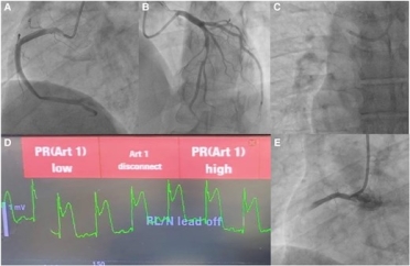
Spot the Diagnosis June 2025 Challenge
#AcuteCVDays may be over, but not the challenging cases – take a peek at the new #SpotTheDiagnosis case. (image in attachment)
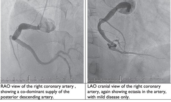
Silent stenosis to sudden shock: the unplanned culprit
What are the next best steps in the management of this patient?
- IV heparin, dual antiplatelet therapy, glycoprotein IIb/IIIa inhibitors, consideration of thrombectomy.
- Vasopressor support, urgent transthoracic echocardiography, preparation for mechanical circulatory support—request cardiothoracic surgeon backup.
- Intracoronary nitroglycerin, serial ECG monitoring, repeat angiographic views, possible PCI.
- Supplemental 100% oxygen, guidewire insertion, catheter aspiration through guiding/microcatheter, intracoronary saline/contrast flushes, intracoronary vasodilators (e.g. adenosine, nitroprusside).
For more updates and new groundbreaking science in area of #cvacute follow us on social media #EHJACVC @EHJACVCEiC.
Spot the Diagnosis February 2025 Challenge
Are you ready for another challenging #SpotTheDiagnosis case?
A mysterious cause of myocardial infarction: look beyond his coronary vessels
What do these images tell you? What is the most likely diagnosis?
- Infective endocarditis
- Polyarteritis nodosa
- Systemic lupus erythematous with fibromuscular dysplasia
- Takayasu arteritis
For more updates and new groundbreaking science in area of #cvacute follow us on social media #EHJACVC @EHJACVCEiC.
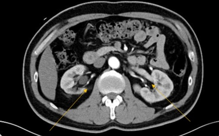
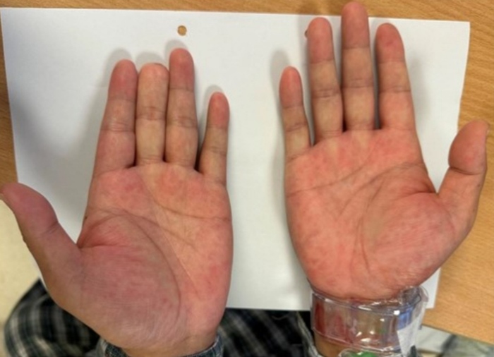
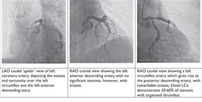
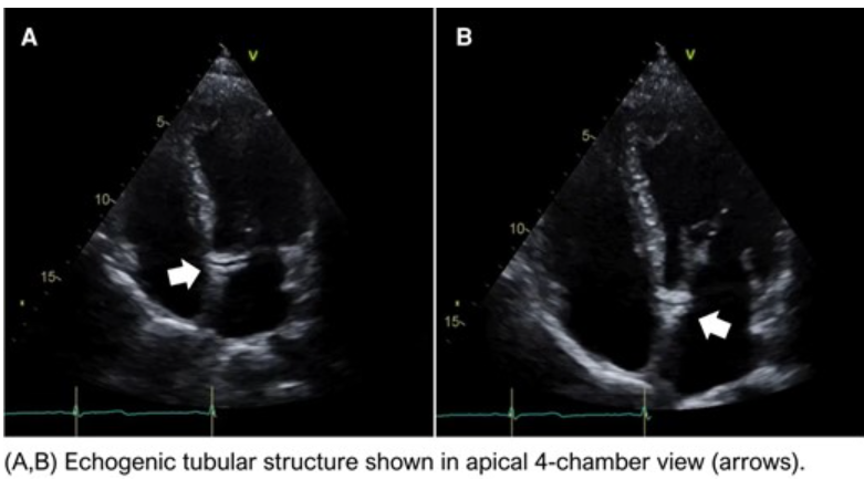
A 44-year-old man with a history of hypertension and smoking arrived at the emergency department with a 2-day history of oppressive chest pain associated with diaphoresis and nausea. Physical examination revealed high blood pressure (170/100 mmHg) and no signs of heart failure. Serial 12-lead electrocardiogram showed dynamic changes of T wave in V5 and V6, and high-sensitivity cardiac troponin levels were increased (443 ng/mL). Point-of-care ultrasound (POCUS) was performed at the bedside showing the following findings:
What is the likely aetiology of this patient’s chief complaint?
- Coronary sinus calcification
- Retroaortic coronary artery
- Septal communication
- Intra-myocardial dissection
For more updates and new groundbreaking science in area of #cvacute follow us on social media #EHJACVC @EHJACVCEiC.
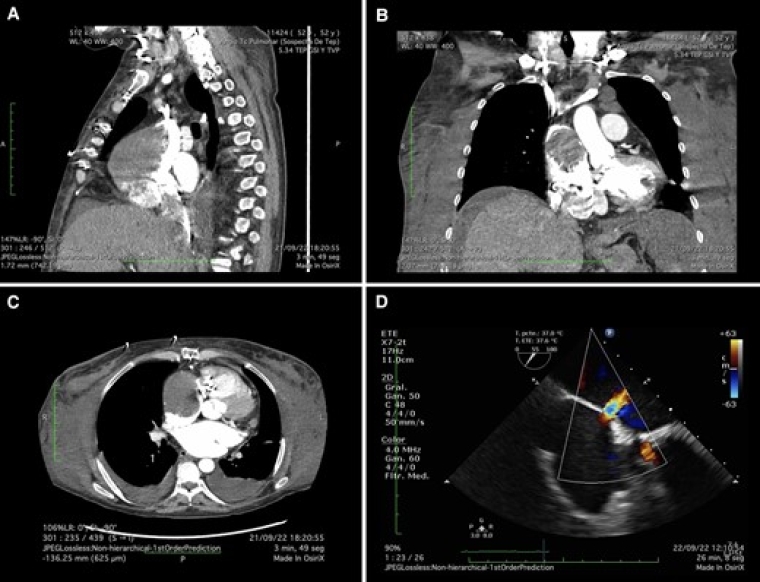
Spot the Diagnosis October 2024 Challenge
October new #SpotTheDiagnosis challenge
What is the cause of refractory hypoxaemia in this patient?
- Pulmonary embolism
- Right to left shunt
- Pulmonary oedema
- Pneumothorax
The answer will be published on our official X account - stay tuned.
For more updates and new groundbreaking science in area of #cvacute follow us on social media #EHJACVC @EHJACVCEiC.
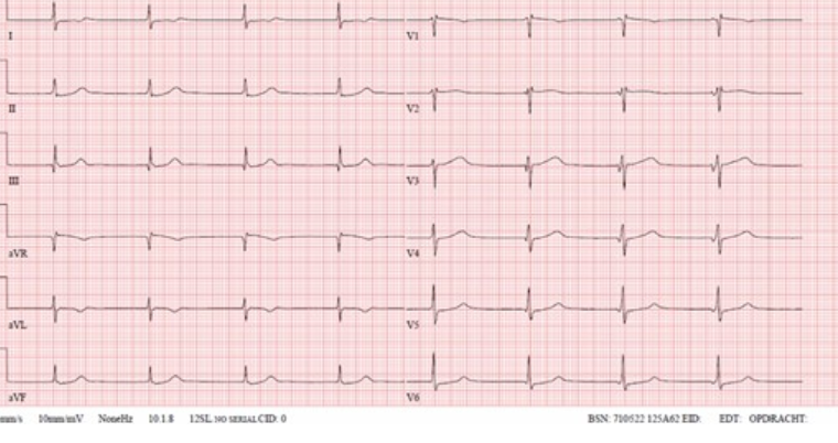
Spot the Diagnosis September 2024 Challenge
What is the most appropriate next step besides continuation of antibiotic treatment?
- Transoesophageal echocardiography
- Lead extraction
- CT scan of the thorax
- Watchful waiting
The answer will be published on our official X account - stay tuned.
For more updates and new groundbreaking science in area of #cvacute follow us on Social Media #EHJACVC @EHJACVCEiC
Why and how to submit in this section
By Frederik H Verbrugge, Deputy Editor EHJ-ACVC
View the latest articles: Question & Answer
Submission link

Follow @EHJACVCEiC
to get the latest updates from the EHJ-ACVC Editor-in-Chief, Pascal Vranckx
Professor Pascal Vranckx
EHJ-ACVC Editor-in-Chief


