In order to bring you the best possible user experience, this site uses Javascript. If you are seeing this message, it is likely that the Javascript option in your browser is disabled. For optimal viewing of this site, please ensure that Javascript is enabled for your browser.
Did you know that your browser is out of date? To get the best experience using our website we recommend that you upgrade to a newer version. Learn more.

Transesophageal 3D echo for heart valve evaluation

An article from the e-journal of the ESC Council for Cardiology Practice

3D transesophageal echocardiography (3D-TEE) has rapidly become the gold standard in the evaluation of every patient with a valve disease. This new technique is both easy to use and results to analyse and provides a new window for heart evaluation, not only for the cardiologist, but also for other medical doctors involved in the management of patients suffering from heart diseases, such as cardiac surgeons.

For the time being, only 3D-TEE probes are commercially available. This probe uses more than 2.500 piezoelectric that allow it to work in two different but complementary ways: 1) as a standard transesophageal transducer, able to obtain 2D, M-mode, Doppler… as when obtaining them with a normal 2D probe (Figure 1). But the same 3D image modalities can also be obtained with a transthoracic 3D probe: x-Plane imaging (two simultaneous planes), Live 3D (real time 3D images), Live 3D Zoom (Figure 2) and 3D Full Volume (four to seven heart cycles are used to obtain a large volume image) and 3D Color Full Volume (several heart cycles are used to obtain a volume with color Doppler information) become possible to acquire. The most frequent limitations of this new system are related to the quality of the transesophageal acoustic window of the patient and to the low temporal resolution it provides.

Aproximate cost of this system is 180.000 euros and the cost of the probe 45.000 euros. Aproximate cost of a 2D TEE probe is 12.000 euros. Cardiologysts are waiting for new devices in the near future and this fact probably decrease the price. Thus, this technique could be affordable for a wide number of  practicionners.

Summarising the most common indications to perform a 3D-TEE, we may say that it is useful in the following situations:

  • Aortic valve evaluation (Figure 3): aortic regurgitation and aortic stenosis assessment (1).
  • Mitral valve evaluation: mitral valve prolapse is one of the most common indications for 3D-TEE (Figure 4) (2,3), but it also can be useful for mitral stenosis evaluation, especially in cases of poor transthoracic echocardiographic window (4).
  • Prosthetic valves evaluation: one of the main indications of 3D-TEE is the evaluation of para-valvular leaks. The information it provides is clearly more accurate that the information provided by 2D echocardiography (5).
  • Left atrium and left atrial appendage assessment (6,7).
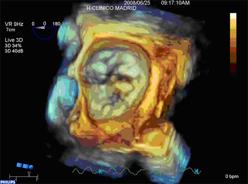
  • Congenital heart disease analysis (Figure 2) (8).
  • Guiding percutaneous interventional procedures (9).
  • Assessment of intra-cardiac masses and tumours (10).

Figure 1: 3D-TEE probe may also be used as a conventional 2D-TEE probe. In this figure we can see a mitral valve prolapse.


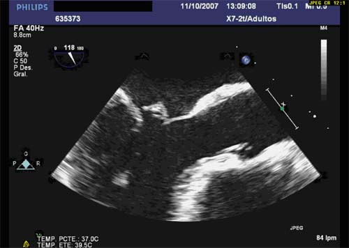
Figure 2: Ostium secundum atrial septal defect obtained by means of 3D-TEE zoom technique. This view was obtained from the left atrium lateral wall. The septal defect can be see as a « black hole » in the image.

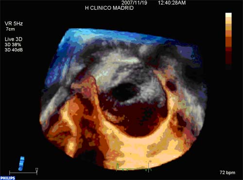
Figure 3: Aortic valve 3D-TEE image. The three aortic leaflets can be seen during systole in this image.

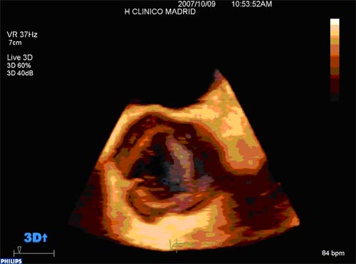
Figure 4: Mitral valve prolapse evaluation by using 3D-TEE imaging. Mitral valve prolapse evaluation is one of the most common indications for transesophageal 3D echocardiography.

Conclusion:

3D-TEE is one of the most significant advances in cardiac imaging from recent years. This new technique is improving the diagnostic accuracy and the clinical management of hundreds of patients all around the world.

References


1. Scohy TV, Soliman OI, Lecomte PV, McGhie J, Kappetein AP, Hofland J, Ten Cate FJ. Intraoperative real time three-dimensional transesophageal echocardiographic measurement of hemodynamic, anatomic and functional changes after aortic valve replacement. Echocardiography. 2009 Jan;26(1):96-9. Epub 2008 Nov 7.

2. Pepi M, Tamborini G, Maltagliati A, Galli CA, Sisillo E, Salvi L, Naliato M, Porqueddu M, Parolari A, Zanobini M, Alamanni F. Head-to-head comparison of two- and three-dimensional transthoracic and transesophageal echocardiography in the localization of mitral valve prolapse. J Am Coll Cardiol. 2006 Dec 19;48(12):2524-30.

2. García-Orta R, Moreno E, Vidal M, Ruiz-López F, Oyonarte JM, Lara J, Moreno T, García-Fernández MA, Azpitarte J. Three-dimensional versus two-dimensional transesophageal echocardiography in mitral valve repair. J Am Soc Echocardiogr. 2007 Jan;20(1):4-12.

3. Langerveld J, Valocik G, Plokker HW, Ernst SM, Mannaerts HF, Kelder JC, Kamp O, Jaarsma W. Additional value of three-dimensional transesophageal echocardiography for patients with mitral valve stenosis undergoing balloon valvuloplasty. J Am Soc Echocardiogr. 2003 Aug;16(8):841-9.

4. Sugeng L, Shernan SK, Weinert L, Shook D, Raman J, Jeevanandam V, DuPont F, Fox J, Mor-Avi V, Lang RM. Real-time three-dimensional transesophageal echocardiography in valve disease: comparison with surgical findings and evaluation of prosthetic valves. J Am Soc Echocardiogr. 2008 Dec;21(12):1347-54. Epub 2008 Oct 10.

5. Jaber WA, White RD, Kuzmiak SA, Boyle JM, Natale A, Apperson-Hansen C, Thomas JD, Asher CR. Comparison of ability to identify left atrial thrombus by three-dimensional tomography versus transesophageal echocardiography in patients with atrial fibrillation. Am J Cardiol. 2004 Feb 15;93(4):486-9.

6. Karakus G, Kodali V, Inamdar V, Nanda NC, Suwanjutah T, Pothineni KR. Comparative assessment of left atrial appendage by transesophageal and combined two- and three-dimensional transthoracic echocardiography. Echocardiography. 2008 Sep;25(8):918-24.

7. Roldán FJ, Vargas-Barrón J, Vázquez-Antona C, Castellanos LM, Erdmenger-Orellana J, Romero-Cárdenas A, Martínez-Ríos MA. Three-dimensional transesophageal echocardiography of the atrial septal defects. Cardiovasc Ultrasound. 2008 Jul 18;6:38.

8. Biner S, Rafique AM, Kar S, Siegel RJ. Live three-dimensional transesophageal echocardiography-guided transcatheter closure of a mitral paraprosthetic leak by Amplatzer occluder. J Am Soc Echocardiogr. 2008 Nov;21(11):1282.e7-9. Epub 2008 Aug 15.

9. Scohy TV, Lecomte PV, McGhie J, Meijer R, Gommers D, Hofland J, Ten Cate FJ. Intraoperative real time three-dimensional transesophageal echocardiographic evaluation of right atrial tumor. Echocardiography. 2008 Jul;25(6):646-9.

VolumeNumber:

Vol8 N°24

Notes to editor


Leopoldo Pérez de Isla, Adriana Saltijeral and José Zamorano
Instituto Cardiovascular Hospital Clínico San Carlos. Madrid. Spain

Correspondence:
 
Leopoldo Pérez de Isla
Unidad de Imagen Cardiovascular
Hospital Clínico San Carlos
Plaza Cristo Rey
28040-Madrid, Spain
Tel: 0034913303290
Fax: 0034913303290
leopisla@hotmail.com

The content of this article reflects the personal opinion of the author/s and is not necessarily the official position of the European Society of Cardiology.