


3D wall motion tracking allows analyzing real myocardial movement in three-dimensions. This technique provides a more complete evaluation of global and segmental left ventricular function.
Myocardial function assessment is an important evaluation in cardiac imaging. Global left ventricular function has been evaluated by different methods. However, segmental wall motion is difficult to assess. Myocardial strain and strain-rate have appeared as quantitative parameters to solve the high variability of left ventricular wall thickening evaluation (1). Until now, both measurements have been obtained from Tissue Doppler Imaging (TDI) datasets, but TDI-based strain and strain-rate have several limitations among which, is of special concern the fact that all the measurements are angle dependent, due to the use of the Doppler effect.
Wall motion tracking
Wall motion tracking is an echocardiographic method which analyzes motion by natural acoustic markers (speckles) in the ultrasonic image. We can say that there is a unique pattern for each myocardial region that can “identify” this specific region. Each speckle can be identified and followed over a number of consecutive frames and the speckles are tracked form one frame to the following one. One of the most interesting advantages of this method is the fact that there is no angle dependency and any kind of Parameters can be calculated in any view. Although 2D-Wall motion tracking is an interesting tool, it has a great limitation: It follows speckle in a 2D plane, but speckle moves in 3 dimensions; thus, only a portion of the real motion can be detected (2-4).
3D wall motion tracking
The main difference between 2D tracking and 3D tracking is that 2D tracking tracks 2 dimensional movements or the projection of 3 dimensional movements into a 2D plane and 3D tracking assesses real movement in 3D space. A global dataset enables to analyze indices such as torsion and opens the door to observe true 3D strain.
When using 2D tracking, a speckle particle can move through the scan plane. Nevertheless, by using 3D tracking, a speckle particle moves through a volume and therefore the vector can be detected along the complete movement. As a result, results for tracking with 3D data are much better. Thus, the detection of the tissue movement is more accurate. The main advantages of the new system are: 1) All vectors of tissue tracking are tracked within the full volume; 2) Faster results are obtained, because all segments are calculated by one analysis step. 3) 3D volumes and ejection fraction are calculated simultaneously. The main indications for this technique are all patients with a special need for a global left ventricular evaluation, such as ischemic patients or patients undergoing resynchronization therapy. The costs of 3D wall motion tracking are comparable to those of 2D speckle tracking except for the time used to perform them: 3D wall motion tracking is less time-consuming.
Figure 1: Wall motion tracking search algorithm: the region of interest is defined in the original frame. In the next frame, the algorithm defines a search region. The search is conducted in all directions for the matching region of interest.
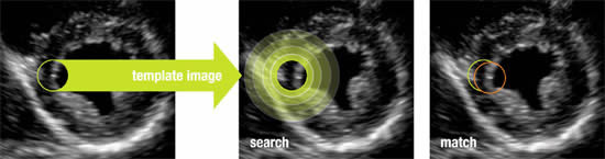
Figure 2: 3D-wall motion tracking display. Left ventricle.
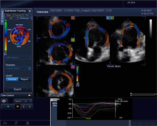
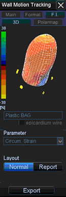
Figure 3: “Plastic-bag” representation of left ventricular circumferential strain obtained from 3D wall motion tracking.
The content of this article reflects the personal opinion of the author/s and is not necessarily the official position of the European Society of Cardiology
Conclusion:
3D-wall motion tracking provides a more complete and probably accurate evaluation of global and segmental left ventricular function. It is a very promising tool to provide new insights in left ventricular function such as real 3D strain, rotation and twist assessment.