


The background of electrophysiology
We have come a long way. Willem Einthoven (1860-1927) needed a 600-pound machine and five operators to capture the first PQRST complex in a human in 1901. The patient had to put two hands and one foot in a bucket with electrolyte solution to obtain a 3-lead electrocardiogram (ECG). Today we take our smartphone and use two fingers to register an ECG [1]. Although electrophysiology may sound like a new field, bioelectricity (in animals) was discovered in 1791 by Luigi Galvani (1737-1798) [2]. Centuries of discourse on anatomy and physiology passed before recording ECGs became feasible.
Pulse and heartbeat
It all started much earlier: in the Chinese and Arab world, pulse measurement had already been practiced for centuries: Wang Chu Ho was said to have authored ten books alone on the pulse by 280 BC [2] and Pien Ts’lo is said to have been the first to have recognised the pulse as a diagnostic toom in the fifth century [3].; The ancient Egyptians described the pulse in the ‘Eber-papers’ [2].
Many centuries later in Europe, Sanctorius Sanctorius (1561-1636) developed techniques for clinical measurements such as pulse rate [4, 5]. In 1717, Marcus Gerbezius (1658-1718) performed a very accurate pulse analysis and described symptoms of bradycardia, probably induced by a complete AV block. This observation was published after his death [6]. Some decades later, Giovanni Morgagni (1682-1771, a pupil of Antonio Valsalva) described the course of a patient with AV block in De sedibus et causis morborum per anatomen indagatis [6]. In the 19th century, anatomy and physiology expanded when more instruments were invented and made available. It became increasingly possible to do research not only in animals, but also in humans.
Origin of heartbeat: myogenic versus neurogenic theory
Physicians and philosophers debated for centuries on the origin of the heartbeat. Claudius Galen (c. 129-c.216 CE, state physician to Marcus Aurelius) wrote that the excised hearts of animals continued to beat for some time, but he did not provide an explanation. Centuries later, the English physician William Harvey (1578-1657) was still not able to explain this in his De motu cordis (1628). In 1651, he held on to Aristotle’s (c. 384-322 BC) theory that blood inside the heart triggers contraction: the myogenic theory.
In the 17th century the neurogenic theory became in vogue. Thomas Willis (1621-1657) stated that the heart was stimulated to contract by a “nervous liquor” that was carried by nerves from the cerebellum to the heart walls [7]. Albrecht von Haller (1708-1777) argued, based on animal experiments, that the heart beats spontaneously. Although a large part of the ‘world’ joined Haller’s theory around 1830, the discussion persisted, especially in France where adherence to the neurogenic theory continued, promoted by Julien Legallois (1775-1814). Around this time, Robert Remak (1815-1865, a pupil of Johannes Müller [1801-1858]) found that frog heart ganglion cells were present in the sinus venosus. Heinrich Bidder (1810-1894) described them at the auriculo-ventricular junction in 1852, and Carl Ludwig (1816-1895) found the same ganglion cells in the interatrial septum [5, 7]. In 1847, Ludwig was the first to outline sinus arrhythmia by recording pulse wave and respiratory patterns simultaneously [8]. Rudolp von Kölliker (1817-1905) and Johannes Müller demonstrated that the heart also produced electricity [9].
Another milestone discovery took place in 1845 when German brothers Ernst Weber (1795-1878) and Friedrich Weber (1806-1871) showed that electrical stimulation of the vagal nerve in animals results in slowing or stopping the heartbeat. They advocated that the nervus vagus was the cause of impulse formation [5]. This again led to new research.
In the 1850s, the nature of the heartbeat was still unexplained. It was Sir Michael Foster (1836-1907) and his Cambridge (UK) group who showed its mechanisms in experiments with jellyfish [7]. He published in 1859 his findings in snails and reported that the heartbeat might be “a peculiar property of the general cardiac tissue” [7]. Foster’s pupil George Romanes (1848-1894) continued research with snail hearts and jellyfish. In the early 1870’s Romanes worked with Walter Gaskell (1847-1914) and Albert Dew-Smith (1848-1903) in Foster’s private lab at his summer home on the Scottish coast [7]. His work laid the foundations for pacemakers and conduction blocks [10]. Foster also encouraged Charles Darwin’s third son, Francis Darwin (1848-1925), to investigate the histology of snail hearts [7]. Darwin observed that there was “a muscular continuity between the auricle and ventricle”. This stimulated Gaskell, who described that the ventricle followed the atrial beat in tortoise hearts in 1883. In parsing this passage, he discovered the ‘block’: the ventricle contracts separately from the atrium [7]. He also mentioned another structure in the auriculo-ventricular groove which possessed rhythmical power. Foster’s experiments led to the confirmation of the myogenic theory in 1870. He researched electrical currents in frogs, and concluded in 1877 that “the beat of the heart is an automatic action” [7]. In 1888, in the 5th edition of his Foster’s Textbook of Physiology, he included the myogenic theory of heartbeat [10].
Still, this evidence was not enough to end the myogenic-neurogenic debate. In 1891, Sir William Bayliss (1860-1924) and Ernest Starling (1866-1927) (Bayliss was married to Starling’s sister) showed that contraction waves in mammals were the same from base to apex as in frogs and cold-blooded animals [7]. Starling is also credited with the Frank-Starlings law. A review by Gaskell in 1900 definitively showed the validity of the myogenic theory [7].
Discovery of conduction system, sinus node and AV node
The Bohemian Johannes Purkinje (1787-1869), who had become professor of physiology in Wroclaw (Breslau) with the support of Johann Wolfgang von Goethe, discovered the fibres that carry his name [11], in 1839. In 1907, Martin Flack (1882-1931) and Sir Arthur Keith (1866-1955) published their discovery of specialised tissue in the sinus venosus in mammals [5, 7].
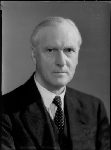
Figure 1. Sir Arthur Keith. © National Portrait Gallery, London
On an earlier boat trip in Scotland, Karel Wenckebach (1868-1940) once suggested to Keith that a histological examination of the sino-atrial junction might be useful. Keith, who had his laboratory at his farmhouse in Kent, was taking a bike ride with his wife on a summer day, while his medical student, Martin Flack (Keith had been looking for an assistant and his grocer suggested his own son Martin) worked on samples from a mole. When Keith returned, an excited Flack reported his discovery: the compact mass of cells reminded Keith of the node of Tawara. In checking all their mammalian specimens, they found the same tissue in all of them [12]. In 1910, Sir Thomas Lewis (1881-1945) described this spot as the ‘pacemaker’ of the heart, which became the sinus node.
By using ECGs, X-rays, sphygmographs and sphygmomanometers - all 19th century inventions that map internal movements of the body [3] - Sir James Mackenzie (1853-1925) and Thomas Lewis were able to bring sinus node research into clinical practice [7, 13], as described in Mackenzie’s “The Study of the Pulse” (1902 [3]).
Basel-born (Switzerland) Wilhelm His Jr (1863-1934), who served in WW1 and described trench fever, discovered the AV bundle in 1893 [5]. Bundle branches were described in 1904 [9]. Stanley Kent (1863-1958) also found around the same time (1893) multiple AV-connections, which later appeared to be features in the WPW-syndrome [14].
German pathologist Karl Albert Aschoff (1866-1942) described characteristic histological lesions in rheumatic myocarditis. He named the reticuloendothelial system [5]. Together with his Japanese pupil Sunao Tawara (1873-1952), he described the AV-node of the conducting system of the heart, in 1906 [5]. Ivan Mahaim (1897-1965), born in Liege, Belgium, studied and worked in Switzerland and became a fellow of Wenckebach in 1926. He did much histological research on connections in the bundle of His. In 1932 he described the Mahaim fibres [8, 15].
Jean George Bachmann (1877-1959) outlined in 1916 in experiments on canines, that clamping the muscular bundle of fibres that connect the atria caused a significant conduction delay. When the sinus node discharges, this bundle spreads the activation to the left atrium, resulting in an almost simultaneous contraction [16]. In 1963, Thomas N. James (1925-2010) described the three pathways connecting the sinus node to the AV node: the anterior, medial and posterior internodal pathways [16]. The anterior pathway displays a second branch at the superior caval vein level to form Bachmann’s Bundle.
Physiology of conduction
Another subject of lengthy debate was how conduction took place. Moritz Schiff (1823-1896) outlined the refractory period in 1850 [7]. Ludimar Hermann (1838-1914) and Julius Bernstein (1839-1966), in 1899 and 1902, respectively, developed the cell membrane theory: local electrical circuit currents as opposed to chemical transmission. In 1939, Alan Hodgkin (1914-1998) provided evidence for local currents in crab and squid [17]. Evidence for the cardiac muscle being a functional syncytium were published in 1952 by Silvio Weidmann (1921-2005), the ‘father’ of cardiac cellular electrophysiology [17, 18]. In the same year, Hodgkin and Andrew Huxley (1917-2012) demonstrated in a squid axon how channels open and close, and how ions travel through these channels. This work was rewarded with the Nobel Prize in 1963.
The discovery of the gap junction in cardiac cells was made by M. Karnokovsky in 1967 [17], while Etienne-Jules Marey (1830-1904) was the first to document ventricular premature beats. He also described the refractory period of the heart, and produced the first graphically recorded ECG in an animal in 1876 [8]. Fun fact, he also invented cinematography.
Pioneers of the ECG
Augustus Desiré Waller (1856-1922) was born in Paris to the famous physiologist Augustus Volney Waller. A.D. Waller was a pupil of Carl Ludwig [8], and worked for years in Scotland before going to the University of London where he became director of the physiology laboratory, studying mainly the electrical aspects of the heart [19]. The electrometer was devised in 1873 by Gabriel Lippman (1845-1921) and in 1887, Waller recorded the first ECG from a human body using the Lippman capillary electrometer and electrode strapped to the front and back of the chest [20]. The Waller ECG consisted of only two reflections: ventricular re- and depolarisation and the P-wave could not be seen. Einthoven witnessed this event. In 1888, Waller recorded an ECG while putting the extremities of his subject in saline jars [19].
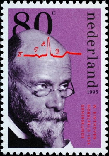
Einthoven started in 1895 with the Lippman capillary electrometer. Later he refined the string galvanometer, invented by Clement Ader (1841-1925) in 1897 [20]. In 1924, Willem Einthoven was awarded with the Nobel Prize for Medicine for “the discovery of the electrocardiogram mechanism”, using the designations P, Q, R, S and T [8, 21]. Why these letters were chosen is still not clear. It was suggested that in choosing letters near the middle of the alphabet, he was leaving ample room for the naming of future waves yet to be discovered. Another explanation recounts that he was influenced by Rene Descartes who had used the P and Q as letters for points on a curve. A third story holds that when he first started to use the capillary electrometer, he designated the curves ABCD, but ended up with PQRST after all his refinements [22].
Figure 2. Willem Einthoven, Dutch stamp to celebrate his Nobel Prize. ©PostNL
Eponyms
Robert Adams (1791-1975) provided the classical description of heart block associated with syncope in 1827, which was named the Adams-Stokes syndrome [5].
William Stokes (1804-1878) described heart block associated with syncopal attacks in 1846, and both paroxysmal tachycardia and Cheyne-Stokes breathing in apoplexy in 1854 [5].
In addition to being a Harvard cardiologist, Louis Wolff (1898-1972) was a concert-quality violinist who enjoyed accompanying his wife, a flutist. He described together with Parkinson and White the WPW syndrome in 1930. He finally met Parkinson face-to-face in 1954 for the first time.
Sir John Parkinson (1885-1976; not to be confused with James Parkinson who named Parkinson’s disease) worked as an assistant to Sir James Mackenzie. The first European Congress on Cardiology in 1952 was chaired by Parkinson.
Paul Dudley White (1886-1973) graduated from Harvard and studied for one year with Sir Thomas Lewis in London. He wrote his first book on heart disease in 1931 and treated US President Dwight D. Eisenhower for his heart attack [5].
Woldemar Mobitz (1889-1951) did his most important work between 1924 and 1928. In 1924 he differentiated two types of second-degree AV block, of which Mobitz type I was identical to the previously described type of second-degree AV block by Wenckebach at the end of the nineteenth century.
Karel Fredrick Wenckebach (1864-1940) was a Dutch physician working in Vienna. He described in 1899 a form of progressive AV block until a drop in ventricular beat occurred, and also wrote about beneficial effects of quinine in arrhythmias [5]. During his stay at the laboratory of Theodor Wilhelm Engelmann (1843-1909), he defined terms such as extrasystole, compensatory pause and concealed conduction, and observed electrophysiology using atrial and ventricular pressure curves of stimulated and isolated shark hearts. In 1891, he went to Heerlen, the Netherlands, to work as a general practitioner. In this work, he was often confronted with irregular heartbeats. He was so fascinated by extrasystoles and compensatory pauses that he went back to Engelmann. In 1899, he described for the first-time regular droppings of heartbeats, which he called “Luciani periods” (Luigi Luciani had described this phenomenon in 1872 in sharks). Wenckebach demonstrated the difference between these observations and extrasystoles. In 1906, Einthoven was able to trace this gradual prolongation of the PR-interval followed by a dropped QRS complex on ECG. Wenckebach also did research on the use of quinine, as he came into close contact with a patient from the colonial Dutch East Indies who used quinine for malaria, and sometimes used a bit more to treat his palpitations.
Anton Jervell (1901-1987) and Fred Lange-Nielsen (1919-1989, also a jazz musician) described in 1957 a Norwegian family with syncopal arrhythmias, QT prolongation and congenital deafness [8].
In 1963, independently of one another, two cases in small children were described by Cesarino Romano (1924-2008) in Italy, and Owen Ward (1923) in Ireland. They reported prolonged QT-interval and syncopal attacks, also in siblings who had already died. The arrhythmia was caused by torsade de pointes (TdP) which were first described by Francois Dessertenne in 1966 [8].
Norman “Jeff” Holter (1914-1983) invented ambulatory electrocardiography in 1947. This first ECG radio transmitter with batteries weighed more than 38 kg. In 1952, it was improved and downsized to 1,18 kg. Later the ‘Holter’ was replaced by a magnetic tape recorder. Holter started his collaboration with Bruce Del Mar to manufacture and market the recorder in 1963 [23]. Today data are stored on flash memory that weighs about 0,07 kg.
Invasive clinical electrophysiology (EP)
Invasive clinical EP is based on the heart catheter technique by Werner Forssmann [13]. EP began in 1967 in Amsterdam, when Dirk Durrer (1918-1984) and Hein Wellens (1935-2020) demonstrated in a patient with Wolff-Parkinson-White (WPW) syndrome that arrhythmias could be initiated and terminated by programmed electrical stimulation (PES). At the same time in France, Philippe Coumel (1935-2004) reported similar findings in a patient with an atrioventricular junctional tachycardia [24-26]. In the early 1960s, Durrer introduced the multi-terminal intramural needle electrode which gave insight into reentry/circus movement as a base for several tachycardias [8]. Wellens described the mechanism of the WPW syndrome [8]. EP was first used to induce ventricular tachycardias (VT) and elucidate WPW mechanisms. Later the effects of pharmacological therapies were studied, as well mechanism of sudden cardiac death [8].
In 1968, Cobb reported that surgical interruption of the bundle of Kent using epicardial mapping could cure WPW-syndrome [27].
Benjamin Scherlag (1932- ) was able to produce consistent His Bundle recordings in humans using a catheter in 1969 [14]. This had been described once before by Giraud in 1960 in a human heart with an ASD [28]. Giraud was the first to describe intracardiac leads in 1960 [8].
Combining interpretations of the 12 lead ECG during arrhythmias and PES data brought new insights. Work by Henry Marriott (1917-2007), as well as by Wellens, led to a deeper understanding of the origin of wide QRS tachycardias [26].
In 1968, Cobb and Sealy et all performed the first surgical interruption in a WPW patient [26].
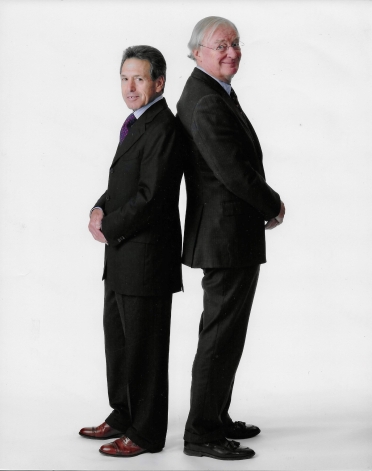
Figure 3. Professor Hein Wellens (right) and Professor Mark Josephson (left). Private collection, Courtesy of Mrs I. Wellens.
Further work by Gerard Guiraudon (1932-2019), Guy Fontaine (1936-2018, who also discovered ARVD) and Mark Josephson (1943-2017) made surgery for VT possible [26]. The first catheter-induced AV block was found by serendipity in 1981 when Rolando Gonzales and Melvin Scheinman reported that a defibrillating electrode accidentally came into contact with an electrode catheter in the His-bundle [27].
Larger series using direct current ablations were performed by Scheinman and Gallagher in 1982 [29]. The first successful radiofrequency (RF) ablation of an accessory pathway was carried out in Munster, Germany in 1987 by Martin Borggrefe et al [29].
In the 1990’s, direct current (DC) ablation was replaced by radiofrequency energy [27]. Later cryoablation also became a possibility.
Mark Josephson was one of the pioneers in catheter-based treatments of cardiac arrhythmias. He was among the first to find the pathogenesis of VT after MI, and developed methods for mapping arrhythmias, which could later be used to ablate them [30]. For 37 years Wellens and Josephson travelled the world to give their famous arrhythmology ECG courses [25].
Surgery for arrhythmias
WPW
The first successful SVT operation was performed in 1968 for WPW [31].
Atrial fibrillation
In 1980, James Cox (1942) was the first to describe left atrial isolation (Maze procedure) for treating atrial fibrillation (AF) [31]. Another option in the early 1980s was surgical AV node ablation with implantation of a ventricular demand pacing (VVI) pacemaker, or transvenously with RF energy. The first surgical test procedure on a patient, which would later become the Cox-Maze procedure, was performed in 1986. In 1987 the first ‘real’ Cox-Maze I procedure was carried out in St Louis, USA [32]. It was modified twice to become the Cox-Maze III in 1995.
The first attempts of surgical ablation (Maze procedure) for AF in humans were reported by Cox in 1989 [26, 33]. In 1994, Swartz reported a catheter-based radiofrequency chronic AF ablation [32].
In 1998 Michel Haïssaguerre (1955- ) published a milestone study using multi-electrode mapping catheters to describe the pattern of the arrhythmogenic-foci-originating AF and ablating with local RF energy in foci in the pulmonary veins [32, 33]. In 1999, the first full Cox-Maze was performed with cryoablation. Cox adapted his technique due to the work of Haïsseguerre which became the Cox-Maze IV in 1999 [32]. This cryosurgical Cox-Maze was also applied as a minimally invasive procedure using a right anterior thoracotomy [32].
Ventricular arrhythmias
In 1959, Orrie A. Couch (1917-2009) reported the first surgical cure of VT by excision of a left ventricular (LV) aneurysm [31. In the 1970s, Josephson and his surgical team introduced subendocardial resection for treating VT’s in patients with ventricular aneurysms and ischaemic heart disease. This was known as the “Pennsylvania Peel” [34]. As this was successful, the step to endocardial treatment was a logical one. In 1983 the first endocardial 300-J DC shock was used as VT ablation. By the end of the 1980s this was switched to RF energy. The 1993 work of William Stevenson (who joined Wellens in Maastricht for six months) on the identification of detailed VT circuit characteristics is still the paradigm for VT ablation guided by circuit mapping [34]. For non-ischaemic VT’s, which are more often localised epicardially, Eduardo Sosa (1942-2020), in 1996, was the first to use percutaneous epicardial access to map and ablate VT’s [34].
In 1999, Carlo Pappone (1961-) improved techniques using a new non-fluoroscopic electroanatomical mapping system [34].
Conclusion
Next year, in 2022, 55 years of electrophysiology will be celebrated. The progress made during these decades has been spectacular, especially in the light of history. It took centuries to understand the structures and functions which form the foundations of current clinical electrophysiology. Hein Wellens put it this way: “There is a golden rule in cardiology that every five years 50% of our knowledge is replaced by new information. This especially holds for cardiac arrhythmology” [31]. While celebrating technical progress, it is good to remember Sir William Osler’s most cherished position: a doctor treats patients, not diseases.