Introduction
Radiotherapy is the use of radiation to kill cancer cells by damaging their DNA. The radiation is most commonly delivered as high energy X-rays but can also be delivered using electrons or heavy particles such as protons. Radiotherapy is often delivered using several shaped beams from different angles in order to maximise the dose delivered to the tumour while minimising the exposure of healthy tissue. In addition, a course of radiotherapy is spread over several days or “fractions” to allow normal cells to repair.
Radiotherapy can be delivered alone, or as an adjuvant treatment in combination with chemotherapy and/or surgery. The treatment is prescribed as a total dose (given in gray or Gy) over a certain number of fractions (e.g., 60 Gy in 30 fractions).
Radiotherapy has undergone several step changes in the last twenty years, driven by advances in optimisation and delivery technology: today’s treatments are highly individualised and tailored to each patient’s anatomy and disease. For example, the introduction of on-board image guidance, where the position of the tumour can be verified each day before treatment, has led to considerably smaller volumes being irradiated, with corresponding oncological efficiency [1]. In addition, an increased awareness of the late side effects of radiotherapy, such as radiation-induced heart disease (RIHD), has led to the development of specific techniques designed to decrease the exposure of the heart.
How radiation affects the heart
Radiotherapy is used in the treatment of several thoracic cancers including breast, lung and oesophagus cancers, as well as some forms of lymphoma. Radiation to the heart activates acute inflammatory pathways causing a chronic pathogenic cascade. The timing and location of the inflammation will determine the cardiac pathology [2,3].
Acute inflammation, at the time of, or immediately after radiotherapy, may cause pericarditis or myocarditis. In the longer term, chronic inflammation can cause widespread pericardial adhesions and thickening, ultimately leading to intractable and inoperable pericardial constriction. Fibrosis and calcification of the aortic root and the aorto-mitral curtain can lead to progressive stenosis of the aortic and mitral valves. Chronic inflammation of coronary arteries leads to accelerated atherosclerosis, with the more proximal sections worst affected.
Radiotherapy: today versus yesterday
Radiotherapy is a loco-regional treatment; therefore, the dose delivered to the tumour gives little information about the dose received by the heart. In a patient treated for mediastinal Hodgkin lymphoma (HL) with 30 Gy, the heart could be exposed to a high dose, or to no dose at all, depending on the exact size and location of the disease within the chest.
There are a number of limitations in the existing literature on cardiac radiation dose that make it difficult to extrapolate published risk estimates to patients treated today. Firstly, in many published studies of RIHD, neither patient-specific heart doses nor doses to cardiac substructures are available. Instead, those studies used very rough measures of cardiac exposure (e.g., “radiotherapy to the chest”). Secondly, there is evidence of different dose responses for different cardiac disease endpoints. Thirdly, there are large variations in practice in modern radiotherapy due to different availability of high-tech equipment and human resources. Finally, the doses of radiation prescribed have changed over time, as have the systemic treatments given alongside radiotherapy.
While the major advances in radiotherapy benefit patients, they make the job of the cardio-oncologist more challenging, as it can be very difficult to gauge how much dose was delivered to a patient’s heart just by knowing their cancer type and/or the region irradiated. The global cardiovascular (CV) toxicity risk of a cancer patient is a consequence of the association of radiotherapy and chemotherapy, particularly anthracycline and trastuzumab. This risk is modulated by the general condition of the patient, the presence of previous CV diseases and of the common CV risk factors such as smoking, hypertension, diabetes and dyslipidaemia. As a result, it is critical to optimise each patient’s cardiac function before the onset of cancer treatment.
In the following sections we review the evolution of radiotherapy and describe modern heart-sparing techniques. This is not intended to be an exhaustive description since radiotherapy techniques keep evolving. However, we hope that this will be informative for cardiologists wondering about the risks of radiotherapy in their patients undergoing cancer treatment today or living with the consequences of an earlier treatment.
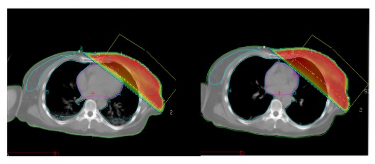
In free breathing (left), a portion of the heart is included within the radiotherapy beams (the dose is displayed as a red colour wash) and the mean heart dose is about 3 Gy. Due to the inflation of the lungs in deep inspiration breath hold (DIBH, right), the heart is pushed away from the chest wall, and out of the beams. Both images represent the same anatomical level within the breast tissue.
The results of the landmark FAST-FORWARD trial [4] show that breast radiotherapy after surgery can be safely delivered as 25 Gy in 5 fractions (over 1 week) compared to 40 Gy in 15 fractions (over 3 weeks). While these shorter treatments are more convenient for patients and highly cost-effective, their effect on the heart is unclear: they are generally safe in the short and medium term, while data on long-term effects are still not available.
The incidence of RIHD in breast cancer survivors is well documented [3]. The rate of major coronary events (myocardial infarction, coronary revascularisation or death from ischaemic heart disease) has been shown to increase linearly with heart dose for patients treated between 1958 and 2001 and is the highest in the first 10 years after radiotherapy [5]. However, a recent analysis of >25,000 patients treated with modern radiotherapy (2008-2016) showed no increased risk of cardiac events in left-sided versus right-sided patients within the first 10 years after radiotherapy [6]. This suggests that heart-sparing approaches have had a considerable impact, though individual patients may still be at risk. Taylor et al suggest that the risk of RIHD with modern radiotherapy is limited in non-smokers [7], emphasising the importance of a healthy lifestyle in this group of cancer survivors.
Radiotherapy for Hodgkin lymphoma
With advances in cancer treatment, HL has gone from being an incurable disease to having an excellent prognosis (>95% survival at 5 years in early-stage disease) [8], due both to the progress in radiotherapy and to outstanding results of systemic chemotherapy.
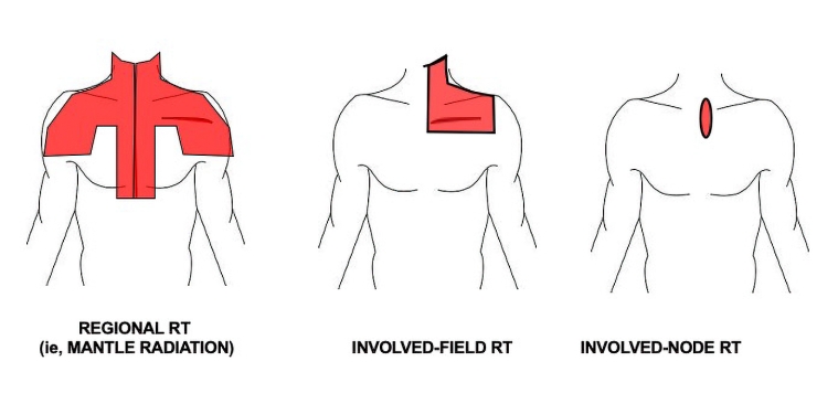
HL is arguably the most dramatic example of radiotherapy evolution. Figure 2 shows the step changes in field size reduction and prescribed doses, due to improvements in image guidance to improve staging and to define the sites of disease (“involved nodes”). The Figure illustrates the differences between mantle fields (“extended field radiation”, which is rarely used today) and involved node or involved site radiotherapy.
Advances and greater individualisation have resulted in a marked decreased exposure of the heart, though the benefit will depend on disease site and location.
Compared with patients with breast cancer, patients with HL may receive a lower dose of radiation but to a larger portion of the heart, depending on the size and exact site of their disease. As with breast cancer, DIBH (see Figure 1) may be used to increase the distance between heart and disease, in addition to advanced delivery techniques, e.g., intensity modulated radiation therapy (IMRT). However, IMRT may spare the heart while increasing the dose to other risk organs (such as the breast), and a balance must be reached between risk of RIHD and risk of radiation-induced second cancers.
Because of their young age at treatment and excellent prognosis, HL survivors form the prototype study group for the late effects of cancer treatments. Van Nimwegen et al have shown that mediastinal radiotherapy increased the risks of coronary heart disease, valvular heart disease, and heart failure for patients treated between 1965 and 1995 using high-dose, extended field radiotherapy, and that this risk was sustained even 40 years post treatment [9]. It is unclear how much modern radiotherapy reduces this risk, but recent research suggests that it should decrease markedly with lower heart exposure [10].
Perhaps even more than in any other patient group, the individualisation of radiotherapy fields means that the risk of RIHD for patients with HL treated today should be evaluated in collaboration with oncologists and following a patient-specific estimate of heart dose.
Radiotherapy for lung and oesophageal cancers
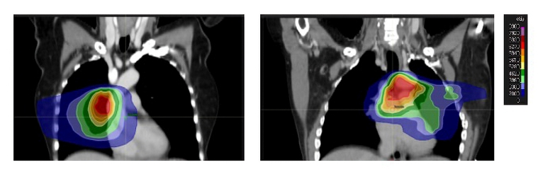
In contrast to the long-standing belief that the survival of patients with lung or oesophageal cancer was too short for RIHD to develop, new research has increased awareness of the link between heart dose and cardiac mortality in these patients. Attempts at escalating the dose of radiotherapy to improve survival have been disappointing and this may be due, in part, to increased heart dose. The radiation dose received by the heart in patients with lung and oesophageal cancer depends on the tumour stage and location. For example, large tumours in the middle and lower lobes of the lung or the lower oesophagus and those involving hilar and inferior mediastinal nodes will result in greater heart dose (Figure 3). Patients receiving radiotherapy to a lower oesophageal tumour have been shown to have an increased risk of cardiac death [11].
Patients with stage 3 lung cancer are treated with higher radiotherapy doses than patients with HL or breast cancer (e.g., 60 Gy in 30 fractions over 6 weeks). Chemotherapy is usually given before and/or during radiotherapy, followed by immunotherapy for a year after radiotherapy in some patients [12]. Patients with stage 3 oesophageal cancer may receive chemo-radiotherapy before, or instead of, surgery to a dose of 45-55 Gy over 20 fractions.
Patients with early-stage lung cancer who are not fit for or do not want surgery will have stereotactic ablative body radiotherapy (SABR), which is high dose, high precision radiotherapy over a short number of fractions (e.g., 54 Gy in 3 fractions on alternate days over 1 week).
These differences in radiotherapy schedules and tumour location will have an impact on the radiation dose received by different cardiac substructures, making it very difficult to define a “safe” heart dose in patients with lung or oesophageal cancer. There is increasing evidence that radiotherapy dose to the right atrium, coronary arteries and aortic valve may be associated with increased risk of post-radiotherapy cardiac events and premature death [13][14]. Moreover, patients diagnosed with lung or oesophageal cancers are often older than breast cancer or HL patients and are at increased risk of cardiac events (independent of radiotherapy) due to smoking, social deprivation and comorbidities. Patients with a history of coronary artery disease (CAD) have an increased rate of major adverse coronary events (MACE) (unstable angina, hospitalisation or urgent visit due to heart failure, myocardial infarction, coronary revascularisation and cardiac death) following radiotherapy for stage 2-3 lung cancer; however, radiation dose to the heart does not significantly increase the incidence of MACE.
Any attempts to reduce the radiation dose to the heart in patients with lung or oesophageal cancer have to be balanced against potential increases in the dose to the lungs. Radiation to the lungs can cause severe, intractable breathlessness and even death due to radiation pneumonitis and lung fibrosis and, currently, reducing lung dose is prioritised over heart dose. Further research is needed to understand how to prioritise heart versus lung sparing in patients with a high burden of comorbidities.
Childhood cancers
Childhood cancer survivors have long been recognised as a high-risk population for late effects of cancer treatment, including anthracycline-based chemotherapy and radiotherapy. The heart may be exposed to radiation in the treatment of lymphoma, Wilms’ tumours, sarcoma, neuroblastoma or following craniospinal irradiation. The largest body of evidence concerning late effects in this patient population comes from large observational studies, e.g., the Childhood Cancer Survivor Study (CCSS, www.ccss.stjude.org). Notably, in a cohort of paediatric cancer patients treated between 1970 and 1999, Mulrooney et al have shown that changes in cancer treatment (e.g., reduced radiation dose to the heart, particularly among survivors of Hodgkin lymphoma) have led to a decreased risk of CAD [15].
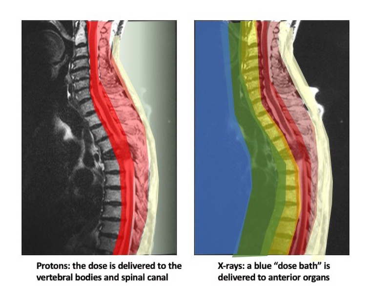
Modern radiotherapy will hopefully reduce this risk even further. For example, children are often prioritised for a new form of radiotherapy called “proton beam radiotherapy” (PBT). Instead of the traditional X-rays, PBT uses the unique physical properties of protons to decrease the amount of healthy tissue being exposed to medium-low dose (see Figure 4). However, it should be noted that PBT is complex; some technological limitations limit its use in some patient groups. It is also considerably more expensive, and less available than conventional radiotherapy. In addition, though it is recognised that PBT reduces dose to healthy tissue, real evidence of the clinical benefits is limited. The results of ongoing trials and multi-national prospective studies are awaited.
Cardio-oncology follow-up for patients treated with radiotherapy
The duration and frequency of long-term cardio-oncological follow-up should be adapted to the level of risk, which will be determined by a patient’s underlying comorbidities, radiation dose to the heart and any adjuvant treatment such as chemotherapy.
For patients receiving mediastinal radiation, monitoring is recommended starting 5 years post treatment and then at least every 5 years thereafter [16]. Patients, even if asymptomatic, should be evaluated for heart failure, valvular disease and CAD. Valvular disease can occur decades after treatment and appear outside of the high radiation dose in patients treated with chemo-radiation. In childhood cancer survivors treated with chest radiation, the International Late Effects of Childhood Cancer Guideline Harmonisation Group recommends lifelong surveillance using echocardiography and at least every 5 years, to monitor for the development of cardiomyopathy [17].
There are currently few guidelines for the cardiac monitoring of patients following lung or oesophageal cancer.
In high-risk patients, such as those with pre-existing cardiac disease or who receive a high heart dose, more frequent monitoring during and immediately after cancer treatment may be appropriate. Cardiac toxicity of radiotherapy may not become apparent until a patient has symptoms and incidence is difficult to determine. For example, high rates of radiation-induced pericardial disease have been reported on follow-up imaging of patients months or even years after radiotherapy for both lung and oesophageal cancers [18,19]. Most pericardial pathology is asymptomatic and resolves without intervention, although some patients can develop severe pericardial pathology; therefore, it is important to continue to monitor these patients. More research is needed to determine the type and frequency of follow-up needed in high-risk patients.
It is important that patients are made aware of the cardiac risk associated with radiotherapy during the consent process for their cancer treatment and encouraged to follow healthy lifestyle recommendations in order to minimise their individual cardiac risk.
Cardiac radiotherapy for arrhythmia: a new indication
SABR is used for the treatment of early lung tumours and is increasingly being explored as a treatment option for refractory cardiac arrhythmias. Small case series in patients with electrical storm or ventricular tachycardia (VT) have demonstrated that a single dose of 25 Gy can reduce the VT burden with minimal short-term side effects [20]. In these series, the radiotherapy target was defined using electrocardiographic imaging, single-photon emission computed tomography (CT) and cardiac magnetic resonance imaging. Cardiac SABR may provide a treatment option for patients with refractory VT and could also give invaluable data on the effect of radiation on the heart. Close collaboration between radiation oncologists and cardiologists is essential in planning, delivering and evaluating this novel treatment.
Conclusion
Radiotherapy techniques have evolved considerably in the last 20 years, resulting in a decreased exposure to the heart in patients treated for HL and breast cancer, and a reduced risk of RIHD compared with the pre-2000 era. However, due to the latency of RIHD, cancer survivors treated pre 2000 may still present with late cardiac toxicity and require active cardio-oncology care. In addition, a subgroup of patients treated today will remain at risk of developing RIHD either because the size and location of their cancer makes it impossible to avoid the heart completely or because of pre-existing conditions/comorbidities. Radiotherapy is a highly individualised treatment; therefore, identification of patients at risk may be better achieved in a multidisciplinary context, including cardiology and medical oncology/haematology as well as radiation oncology professionals.


