Background
Chaotic electrical and mechanical activity in the heart causing impaired contractility defines atrial fibrillation (AF). Prevelance in the developed world is 1.5–2% and with it, ensue hemodynamic and thromboembolic events that are responsible for significant morbidity and mortality (1-2).
Catheter techniques are used for ablation of usually existing, so-called fast or slow AV nodal pathways for the treatment of tachyarrhythmias. The ablation of not all the atrioventricular (AV) node but part (one or more than one) of AV pathways is referred to as AV node modification - i.e modification of AV conductivity.
The atrioventricular (AV) nodal dual-pathway theory holds that AV nodal pathways associated with chronic atrial fibrillation are present on rhythmogram (3). Meanwhile, more than one third (38.8%) of patients with permanent atrial fibrillation in previous studies had two, three, or four AV nodal pathways (4,5). We suggest that the number of AV nodal pathways discovered in our study subjects represents the number that would be found in the atrial fibrillation population at large (usually two or more) (5,15). Furthermore, we suggest that the number of peaks on RR interval histogram shows the number of AV pathways, and allow us to make a decision regarding recommendation for treatment- ablation or modification of the AV node.
1 - Computer assisted analysis method of RR interval histogram to identify underlying AF pathomechanisms
Analysis of heart rate variability, RR interval histograms and Lorenz plots are among the methods to assess RR intervals however, to identify the underlying atrial fibrillation pathomechanisms and predict the best therapy and its efficacy, various methods have been suggested:
- Surface electrocardiogram (13): The monitoring of ECG in patients during atrial fibrillation show multiple AV nodal pathways and 6.4% of all patients with atrial fibrillation, to obtain heart rate reduction, would benefit from ablation of AV nodal pathways with shorter refractory periods (i.e. atrial node modification) (3,4).
- Bimodal histogram: other authors have hypothesised that a bimodal histogram indicates dual AV nodal physiology and predicts a better outcome after AV node slow pathway conduction modification rather than ablation in chronic atrial fibrillation (5-8).
- Computer assisted analysis method of RR interval histogram: the purpose of our data collection was to investigate the morphophysiology of AV node by using the R-R intervals histogram analysis method. Our work that lasted 11 years and ended in 2009, was to 1) use a non-invasive as opposed to an invasive electrophysiological study computer assisted analysis method of RR interval histogram, 2) examine permanent atrial fibrillation patients before and after restoration of sinus rhythm by radiofrequency ablation and 3) determine the presence of complex structure/morphology of AV nodal pathways in these patients. The obtained data was used to select patients who are considered candidates for radiofrequency modification of AV nodal conduction as opposed to total AV nodal ablation.
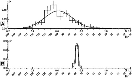
Our study included 64 patients with AF only suitable for recording R-R intervals histograms and its analysis (not treatment). Recruited patients were already treated for AF in Kaunas Clinics. Sinus rhythm was restored by catheter ablation in 19 patients. Coronary artery disease was commonly associated with the atrial fibrillation; concomitant disease in 67% of patients was hypertensive heart disease. In all patients with atrial fibrillation and patients with restoration of sinus rhythm by catheter ablation, 500 RR intervals were recorded in each position: supine during an active orthostatic test, upright, and again in the supine position and after exercise tests (9). Following successful ablation using radiofrequency energy, the examination of patients was repeated the next day (Fig. 1). The long succession of RR intervals was analysed using a von Neuman method (10) and computerised rhythmogram analysis for diagnosing atrial fibrillation (11, 12). In all histograms between 0.2 s and 1.2 s, the RR intervals were classified into 0.025 or 0.05 s wide subgroups. According to the number of peaks, three types of RR interval histograms were distinguished: (a) unimodal, (b) bimodal, (c) and polymodal. The number of peaks in each patient’s RR interval histogram was measured.
2 - Two or more peaks on the RR interval histogram with atrial fibrillation suggested the presence of multiple AV nodal pathways (one peak = one pathway)
The mean age of patients was 64+-9 years, 26% of them were female. The duration of chronic atrial fibrillation was 8.8+-7.3 years. Computer-based analysis of the histograms in patients with atrial fibrillation showed that a single peak was present in 45% of patients, 40% of patients had two peaks, and 15% of patients had three or more peaks. After radiofrequency ablation, ventricular rate (before ablation RR1=0.79+-0,14s, after ablation RR2=0,99+-0.12s) and general heart rate variability (before ablation +-1=0.16+-0.03s, after ablation +-2=0.03+-0.01s) decreased with loss of the peak of short RR cycles after ablation. Presented values are means ± SD. In patients with atrial fibrillation with bimodal and polymodal histograms, the number of peaks decreased in all cases after successful radiofrequency catheter ablation (Fig. 2). Therefore our data showed that two or more peaks on the RR interval histogram with atrial fibrillation suggest the presence of multiple AV nodal pathways.
We compared our results from derived from RR interval histogram computer-based analysis with data from other studies that included patients with atrial fibrillation with unimodal, bimodal, and polymodal RR interval histograms (3, 5, 13, 14). The number of peaks differs in various studies. Our investigations showed that radiofrequency catheter ablation is an effective and fast method for treatment of patients with chronic atrial fibrillation. According to our and literature data (8), this method may prove to be useful in selecting patients with atrial fibrillation who are considered appropriate candidates for radiofrequency modification of AV nodal conduction as opposed to AV nodal ablation. The patients with bimodal or polymodal pattern of RR interval histogram may be more suited for radiofrequency catheter modification of the AV node. Patients with a unimodal pattern may be inappropriate candidates for radiofrequency modification of the AV node and should be referred primarily for AV junction ablation and pacemaker implantation (9).
Conclusions
- Analysis of RR interval histogram in patients with permanent atrial fibrillation showed the different number of peaks on the RR interval histogram.
- Bimodal RR interval distribution during permanent atrial fibrillation in our group of patients confirm the presence of dual AV nodal physiology: in studied patients we registered more than one peak on RR interval histogram and confirm the relation the number of peaks with the number of AV nodal pathways.
- The RR interval histogram computer-based analysis method used in patients with atrial fibrillation as opposed to intracardial electrophysiological study is non-invasive and is useful for explorations of atrioventricular nodal physiology, morphology, and clinical aspects and in view of choosing a treatment method.
Fig. 1 A: RR intervals during atrial fibrillation in supine position, B: RR intervals in supine position after restoration of sinus rhythm by radiofrequency ablation. Nr – serial number of RR interval. S – time scale in seconds.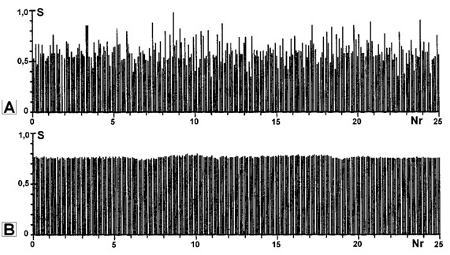
Fig. 2 A: Bimodal RR interval histogram during atrial fibrillation, B: Unimodal RR interval histogram after restoration of sinus rhythm by radiofrequency ablation. The curves on A and B represents Gaussian and experimental RR intervals distribution modes. Hr – heart rate, S – time scale in seconds.