The human aortic wall is composed of three layers and different cell types: the intima, media and adventitia composed of endothelial cells, vascular smooth muscle cells (VSCMs), fibroblasts, mesenchymal stem cells, pericytes and immune cells. Overall, VSMCs are considered the most important cell type in the pathogenesis of aortic aneurysm, although the role of other cell types should not be disregarded (Mackay et al, 2022). In this commentary, we will focus specifically on some new VSMC-related insights.
Historically, VSMCs were believed to switch between a contractile and a synthetic state in response to vascular injury and to withstand the dynamics of blood flow and stress on the aortic wall. Contractile VSMCs are characterized by high expression of contractile genes, whereas synthetic VSMCs express the contractile genes to a lower extent and display increased proliferation, migration, and extracellular matrix production. Dysregulation of this so-called phenotypic switch is hypothesized to be a key process in aortic aneurysm formation. Recent single-cell transcriptomic studies have demonstrated that the different cell types in the aortic wall are far more dynamic than previously thought.
In a single-cell transcriptomic study of ascending aorta and aortic root samples (n=3 per group) of a Marfan mouse model (Fbn1C1041G/+) and one human MFS patient, a distinct modulated VSMC cluster (not present in controls) was identified (Pedroza et al, 2020; Pedroza et al, 2021). These modulated VSMCs had high extracellular matrix (ECM) modulation, collagen synthesis, adhesion and proliferation. In another single-cell RNA-sequencing study of human aneurysmal ascending aortic tissue (n=8, compared to 3 controls), five different types of VSMCs were characterized: contractile VSMCs, stressed VSMCs, two subtypes of proliferating VSMCs (high proliferation gene expression but no migrational or ECM production signature) and fibromyocytes (or modulated VSMCs: only ECM production and no migration or proliferation) (Li et al, 2020), supporting the plasticity and divergent phenotypic nature of VSMCs. The same group also reported on single-cell analysis of ascending aortic aneurysm tissues from three MFS patients (compared to four controls). In addition to a continuum between fibroblasts and VSMCs, they identified a dominant cluster of de-differentiated, proliferative VSMCs in MFS aortic tissue (Dawson et al, 2021). Whether these clusters are identical to the modulated VSMCs from the Pedroza studies will require further study.
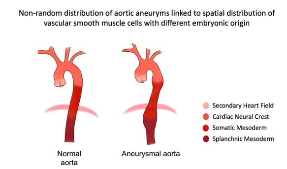
An additional layer of complexity is the spatial difference in embryonic origin of the VSMCs in different anatomical regions of the aorta (see figure). Already in 2019, Elena Gallo-Mc Farlane very elegantly demonstrated in an Loeys-Dietz syndrome Tgfbr1-knock in mouse model that secondary heart field-derived (SHF-derived), but not neighboring cardiac neural crest-derived (CNC-derived), VSMCs showed impaired Smad2/3 activation in response to TGF-β (Gallo Mc Farlane et al, 2019). Similar observations were made when comparing SMAD3-/- (c.652delA) human induced pluripotent stem cell (iPSC)-VSMCs derived from cardiovascular progenitor cells or neural crest cells (Gong et al, 2020). A recent study by Sawada et al, confirmed the critical contribution of second heart field-derived cells to angiotensin II-mediated ascending aortopathies (Sawada et al, 2022).
Overall, the elucidation of the heterogeneous nature of the vascular smooth muscle cells in the aortic wall based on lineage tracing and single-cell transcriptomics has advanced our understanding of the complex biology underlying the development of aortic aneurysmal disease.