- What are blood thinners?
- Vitamin K antagonists
- What is important when using vitamin K antagonists?
- What are precautions while using vitamin K antagonists ?
- Novel (or Non vitamin K) Oral AntiCoagulants (NOAC)
- Oral anticoagulants side effects
- Assessing your risk of bleeding with blood-thinning medicine
- Counteracting oral anticoagulation
- Left Atrial Appendage (LAA) Occluders
- Rhythm control versus rate control
- Drugs used to reduce the episodes of atrial fibrillation (rhythm control):
- Drugs used to slow the heart rate (rate control):
- Electrical cardioversion
- Catheter Ablation of atrial fibrillation
- Pacemaker Implantation combined with atrioventricular node and His Bundle ablation
Treatment of atrial fibrillation may consist of drugs for the arrhythmia itself (anti-arrhythmic drugs), interventional therapy or drugs for treating risks associated with atrial fibrillation (e.g stroke with blood-thinning medication, ‘oral anti-coagulants’).
What are blood thinners?
There are different types of blood thinning drugs. The anticoagulants prescribed in patients with atrial fibrillation are: vitamin K antagonists and the novel oral anticoagulants or NOACs. These latter are also called “direct oral anticoagulants: (DOAC’s) or non-vitamin K oral anticoagulants (NOACs).
- If you need surgery, including dental, let the surgeon know that you are using blood-thinning drugs.
- Using a blood-thinning medicine it is important to you check with a doctor or pharmacist every time you are prescribed a new medication, or when you buy medicines to make sure that they are all compatible. Especially with other blood-thinning drugs, like aspirin.
- The final decision what type of anticoagulation a patient should get, will be taken by the doctor. Some patients might not be good candidates for a NOAC while in other patients one of the NOAC might even provide a greater reduction in stroke risk with comparable or even less risk of intracranial bleeding (bleeding in the brain).
Vitamin K antagonists
Warfarin (coumarin), Acenocoumarol, Phenprocoumon, Fluindione
Vitamin K antagonists are the oldest and worldwide most used anticoagulants. They reduce the ability of the blood to form clots. These blood clots are formed by blood cells and other material to form a solid mass inside the blood vessel. Blood clotting factors are needed for this process. Vitamin K antagonists prevent some of these clotting factors from working, making it more difficult to make clots. This is also called: “making the blood thinner”. Because of this effect of vitamin K antagonists on the clotting factors, they are very effective in reducing the risk of stroke.
What is important when using vitamin K antagonists?
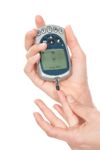
Regular blood tests are required to assess the blood-thinning effect. This is measured by the so called “INR” or international normalized ratio. Depending on this measurement the dosage of the drugs will be adapted on an individual basis. It is of utmost importance to perform this blood test regularly to make sure that the dosage you are using is correct and you are not at risk. The dosage used can differ from person to person and from period to period within one person.
What is the INR and how is it measured?
Normally in persons not taking anticoagulants, blood clots with an INR of around 1.
To reduce the risk of stroke in patients with atrial fibrillation the INR should be between 2 and 3.
You should not stop taking vitamin K antagonist without consulting your doctor.
What are precautions while using vitamin K antagonists ?
Avoid too much alcohol.
Foods that contain high amounts of vitamin K can make the blood-thinning less effective. If your diet is high in vitamin K you will probably need to take more of the blood-thinning drug. The attached table details foods which are low and high in vitamin K (see Table 1 below). Particular care is needed with herbal medications. Always consult your doctor or pharmacist before starting any herbal medicines.
Table 1.
| Foods that are LOW in vitamin K | Foods that are HIGH in vitamin K | |
| Artichoke Asparagus Banana Beans Beetroot Carrots Cauliflower Celery Cilantro Corn Aubergine Green beans Green peppers Mushrooms Onions Parsnip Peas Peeled cucumber Potato Pumpkin Radish Red cabbage Summer squash Sweet Potato Tomato Turnips |
Avocado Amaranth leaf Broccoli Brussel sprouts Cabbage Canola Oil Chayote Chives Coleslaw Collard greens Coriander leaf Endive Fish packed in oil Garbanzo beans Green cabbage Green tea Kale Kiwi Lentils Lettuce Liver Mayoonaise Mint leaf Mustard greens Natto (fermented soybean) Nightshade leaf |
Okra Olive oil Parsley Purslane Seaweed Soybean oil Soybean Spinach Swiss chard Tea from Tonka beans Toumayo Turnip greens Tziton Vegetable drinks (V8) Watercress Wheat grass powder |
Novel (or Non vitamin K) Oral AntiCoagulants (NOAC)
Dabigatran (Pradaxa or Pradax), Rivaroxaban (Xarelto), Apixaban (Eliquis), Edoxaban (Lixiana or Savaysa)
In patients, treated with a NOAC, the kidney function has to be monitored by the doctor. If the kidney function decreases too much the doctor may need to lower the dosage or even stop the blood-thinning medicine.NOACs have been introduced in 2008-2009 and since then successfully used and tested in many thousands of patients with atrial fibrillation to prevent strokes. The drugs directly counteract one of the clotting substances with similar and maybe even better stroke prevention compared to a vitamin K antagonist. They can be used without controlling the INR and are not influenced by food, other drugs and alcohol. Dabigatran and apixaban are taken twice a day and rivaroxaban and edoxaban can be taken once daily. It is extremely important that a NOAC is taken every day as prescribed by your physician.
Oral anticoagulants side effects
The most common side effect that occurs with all blood thinning medications is bleeding. In most cases, bleeding is not serious. Minor bleeding, such as bruising or a minor nosebleed can occur. About 1-2% of people on blood thinners will develop more serious bleeding which may require a blood transfusion and interruption of the blood thinning treatment. The most serious bleeding side effect from blood thinning medication is a bleed into the brain, known as an "intracranial haemorrhage".
If you are concerned about the risk of bleeding associated with your blood-thinning medication, please discuss this with your doctor. Your doctor will assess your individual risk of stroke and weigh up your risk of bleeding with a blood-thinning medicine.
Your doctor can calculate your risk of bleeding before you are offered one of the blood-thinning medicines using a risk score. One example of a commonly used bleeding risk score is the HAS-BLED score.
Assessing your risk of bleeding with blood-thinning medicine
Click here for an online calculator
Counteracting oral anticoagulation
When serious bleeding occurs in patients treated with oral anticoagulants an antidote might be warranted.
The antidote for a vitamin K antagonist is vitamin K itself. The effect of the vitamin K has to be checked several hours later by controlling the INR. If this is still too high another dose of vitamin K has to be given. Additional treatment can include blood clotting products if warranted.
For one of the NOACs: dabigatran (pradaxa) an antidote is also available. It is called idarucizumab (praxbind) and has become clinically available in 2015. One dosage of this antidote is sufficient to completely counteract the effect of dabigatran and normal blood clotting is restored. For the other NOACs there are no clinically available antidote, but will most probably become available in the near future. As with the vitamin K antagonists, blood product may be given to counteract the blood thinning effects of these agents if required.
Left Atrial Appendage (LAA) Occluders
atrial fibrillation results in ineffective atrial contraction, poor atrial blood flow and stasis. This stagnation of blood is thought to favour the development of clots, typically within a finger like pouch of the left atrium, called the left atrial appendage. These clots can result in blockages of critical arteries, e.g. causing strokes. Because most clots form within the left atrial appendage, excising or obliterating the appendage has been performed, principally to reduce the risk of embolic strokes.
For patients who cannot take oral anticoagulants because of bleeding risks, the physician can occlude or obliterate the left atrial appendage through a puncture of the femoral vein in the groin. A plug like device is delivered in collapsed form into the appendage and expanded so as to completely occlude and even obliterate the cavity of the left atrial appendage. The procedure is performed under sedation or general anesthesia. Combination medicines for preventing clotting are necessary for the first few weeks to 3 months; afterwards they can be reduced to a single medicine. Recent studies have found that this procedure provides protection from stroke similar to that with classical oral anticoagulation (with vitamin K antagonists), although there may be immediate complications from the implant procedure.
Rhythm control versus rate control
Rhythm control implies that you and your doctor has decided to obtain and maintain normal sinus rhythm. When this strategy fails or is impossible the presence of atrial fibrillation is accepted or inevitable, it might be important to lower the heart rate. This is called rate control.
Drugs used to reduce the episodes of atrial fibrillation (rhythm control):
The drugs used to reduce or suppress episodes of atrial fibrillation are called "anti-arrhythmics". The most commonly used anti-arrhythmic drugs are:
- flecainide
- amiodarone
- propafenone
- disopyramide
- dronedarone
- sotalol
Anti-arrhythmic drugs modify the electrical activity of heart cells. The generation and transmission of electric signals coordinates the pumping activity of the heart muscle. Taking anti-arrhythmic drugs may help to reduce your chances of relapsing back into atrial fibrillation, but these drugs do not guarantee that you will not experience episodes of atrial fibrillation. Sometimes you may have to try several anti-arrhythmics before you find the one that suits you best and some of the anti-arrhythmic drugs are not suitable for all patients. Your doctor will decide on the most appropriate anti-arrhythmic drug for you depending on the other medical conditions you have, for example, if you have ischemic heart disease (narrowing of the arteries in the heart) or heart failure (poor pumping of the heart). Some anti-arrhythmic drugs such as amiodarone can have serious side effects, so your doctor will monitor you regularly to ensure that it is safe for you to continue taking this medication. For safety reasons, it may be necessary to take a small blood sample to monitor the drug. Your doctor will help to choose the best anti-arrhythmic drug for you and discuss the benefits and possible side effects in more detail with you.
Drugs used to slow the heart rate (rate control):
There are several drugs, taken as tablets, that can help to slow the heart rate down in people with atrial fibrillation and your doctor will choose a drug that fits best.
The main types of medicines that are commonly used for slowing the heart rate are:
- Digitalis medicines
- Beta-blockers (for example: bisoprolol, metoprolol, atenolol)
- Calcium channel antagonists (for example: diltiazem, verapamil)
For many patients with atrial fibrillation one tablet is enough to slow the heart rate down sufficiently but some patients may need to take two or more types of tablets in order to control their heart rate.
Digitalis medicines, such as digoxin have been used for over a hundred years and can slow down even a very fast heart rate. These medicines are very effective especially if you are not very physically active. However they may be less effective during physical activity. This is one of the reasons why another class of drugs, called beta-blockers (e.g bisoprolol or metoprolol) is often used in younger and/or more active patients. Beta-blockers are not suitable for everyone, particularly if you have asthma, and in such cases calcium channel blockers, such as diltiazem or verapamil may be the best option. Your doctor will take into account all of your individual circumstances when deciding on the best option(s) for medicines to help control your heart rate for you.
Electrical cardioversion
If you have an episode of symptomatic atrial fibrillation, which does not respond to anti-arrhythmic drugs, your doctor may suggest that you have a procedure called an electrical cardioversion. This procedure aims to restore your heart rhythm back to a normal rhythm (sinus rhythm). This procedure is usually planned in advance and involves delivering a controlled electric shock to your chest targeting the heart. The electrical impulse is strong enough to briefly stop any electric signals generated by the heart and to let the hearts natural pacemaker: “the sinus node”, regain control over the rate of your heart.
Electrical cardioversion is done in the hospital using a machine called a defibrillator, which is essentially a powerful computerized battery. The electric shock is delivered through two large stickers or paddles (electrodes) attached to the chest. The stickers are usually placed at the front and back of the chest or on the right and left sides of the chest.
Before the cardioversion you will be given an injection (anesthetic) to help make you feel drowsy as this procedure is uncomfortable. This way you will not feel anything during the cardioversion itself. If the first electric shock fails to bring your heart back into a normal rhythm (sinus rhythm) then another electric shock will be attempted using a slightly stronger electrical impulse. You should not feel any pain during the cardioversion but often the areas under the electrodes can be a bit sore for a day or two after the procedure.
You should be aware that even after a successful cardioversion (your heart rhythm has returned to normal sinus rhythm); it is possible that atrial fibrillation returns. This happens in about half of the patients during the first year after the cardioversion. The chance of atrial fibrillation returning depends on many factors but this is more likely if you have other heart problems (including high blood pressure) and if you have had atrial fibrillation for more than 1 year.
It is also possible to acheive a cardioversion using certain medications, which are used to control the rhythm. This is called pharmacological cardioversion as medication is used instead of the electric shock to try to restore your heart rhythm to normal. This procedure is also done in hospital. You will be given this medication through a drip in your arm and your heart rate will be monitored continuously during the procedure.
Before you have a cardioversion, whether this is an electrical cardioversion or a pharmacological cardioversion, you will need to take an anticoagulant, for at least 3 weeks before the procedure. Otherwise, a transesophageal echocardiographic examination has to rule out any blood clots within the heart before the cardioversion. You will also need to continue your blood thinning medicine for at least 4 weeks after the cardioversion procedure to reduce your risk of stroke. Depending on your overall risk of stroke your doctor may ask you to continue taking a blood-thinning medicine for the rest of your life.
Catheter Ablation of atrial fibrillation
Ablation therapy (Pulmonary Vein Isolation, PVI)
The pulmonary veins are vessels connecting the lungs to the left atrium that allow oxygen-enriched blood to return to the heart. Tongues of atrial muscle extend for a short distance (up to about 5 cm) from the body of the left atrium into the pulmonary veins. In people with atrial fibrillation, these muscle extensions have been found to exhibit electrical activity that is rapid and not controlled by the normal activity of the heart. These abnormal electrical discharges, as a result of complex interactions, produce rapid (400-650 per minute) and irregular electrical activity and correspondingly feeble mechanical contractions in the atria.
The spread of abnormal electrical activity originating from the pulmonary veins can be stopped by irreversibly inactivating/killing the cells at the junction of the PVs with the left atrium so that they cannot conduct electrical signals. Inactivation of the cells at the junction of the PVs can be achieved by either heating the cells (to more than 55 degrees but below 80 - 90 degrees centigrade) or cooling the cells (to below -50 degrees centigrade) or by cardiac surgery.
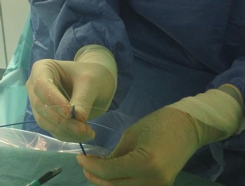
Through small punctures at the groin, the physician introduces multiple catheters into the femoral vein, and advances them under X-ray visualisation to the right atrium. To reach the left atrium, the physician uses a long needle to puncture the wall separating the right and left atria, and then introduces the catheter(s) into the left atrium.
Radio-frequency catheter ablation (RFCA)
The commonest technique uses a high frequency alternating current (about 550 KHz) delivered by a flexible catheter with a metal electrode at the tip. Current passage heats a small tissue volume to more than 55 degrees centigrade depending upon current intensity and tissue contact. Each small volume of inactivated (coagulated) tissue is a ‘lesion’. A series of contiguous, full thickness lesions in a circular design at the junctions of the right and left sided pulmonary veins results in an electrical barricade insulating the pulmonary veins from the left atrium without damaging the physical integrity of tissue. This is pulmonary vein isolation by radio-frequency catheter ablation (RFCA).
During this procedure, the physician introduces two catheters into the left atrium, one only for recording signals from the target pulmonary veins and the other for delivering RF current at a desired location as well as recording from it. The procedure is guided by a reconstruction of the anatomy and shape of the pulmonary veins and left atrium.
The resulting electrical isolation is verified by the stable disappearance of electrical signals in the pulmonary veins and the catheters are then removed.
Cryoballoon Ablation
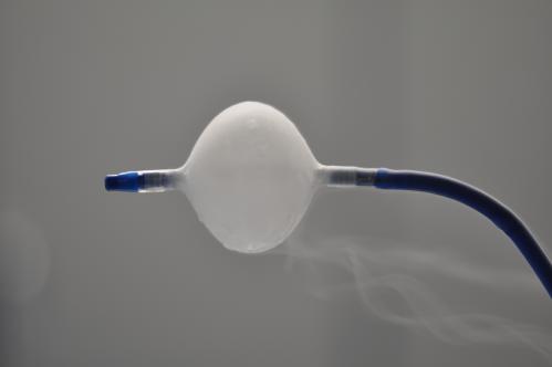
For cryoballoon pulmonary vein isolation , a special deflated balloon (cryoballoon) is introduced into the left atrium then inflated and placed snugly at the mouth of the pulmonary veins. A pressurised refrigerant is injected into the balloon allowing it to expand, and in so doing, it absorbs heat, producing very low temperatures, adhering to and freezing the tissue in circumferential contact with the balloon perimeter. After sufficient freezing, the balloon is warmed, deflated and moved to another pulmonary vein and the ablation repeated. When the balloon is placed in contact with the junction of the pulmonary veins with the left atrium, the resulting circular cryolesion produces pulmonary vein isolation.
Irrespective of the technique used, pulmonary vein isolation is performed with the patient either completely anesthetised and artificially ventilated or powerfully sedated but breathing spontaneously. After the procedure, a compression bandage is applied to the groin and followed typically by a discharge home the following day. After discharge, you can rapidly resume a normal life after the first few days. You will typically need to continue anticoagulation drugs (to prevent clots) for at least 8 weeks after the procedure. Often, some additional medicines are prescribed to control the heart rhythm and rate.
Complications of Pulmonary Vein Isolation
The commonest complication is bleeding from the puncture site in the groin, with a resulting collection of partially coagulated blood under the skin or deeper in the tissue producing a lump, nodule or bruise and frequently tender or painful. Manual compression and bed rest normally can stop the bleeding and the collection progressively disappears over the next few weeks. If at the puncture site, a lump appears suddenly or enlarges or becomes painful, the patient should consult his/her physician.
A clot can be dislodged from within the left atrium and migrate to any of the systemic arterial blood vessels e.g. to the brain, the eye, the limbs, the kidneys etc. thereby blocking the delivery of oxygen, and producing tissue death. This can result in a stroke or damage to the eye, limbs or kidneys. Typically, correct anticoagulation drug use for 3-4 weeks leading up to the procedure, as well as throughout and after the procedure have greatly reduced its incidence to less than 1%. Most physicians also perform special ultrasound imaging of the left atrium from within the esophagus (trans-esophageal echocardiography) to be sure there is no clot before the procedure. A clot can be dissolved in certain instances by a clot dissolving medicine or the blocked artery can be reopened by passing a balloon through the clot, thus restoring blood supply.
Internal bleeding can occur, typically in the sac around the heart, resulting from damage to the wall of the heart – in about 1-2%. This can result in a state of shock with the heart unable to deliver sufficient blood to the body. In most cases, a needle puncture from just under the breast bone can be used to introduce a tube-catheter into the sac around the heart and drain the blood. This is usually enough to restore normal circulation otherwise surgery may be required.
Damage to surrounding tissue outside the heart can occur including to nerves and the oesophagus. The nerve to the diaphragm (an important muscle for breathing) can be damaged, typically more frequently by cryoballoon ablation, resulting in shortness of breath but usually recovers spontaneously within weeks to months. Nerves to the stomach can also be damaged, typically resulting in bloating and abdominal fullness but again this often recovers within weeks.
The oesophagus which is located just behind the back wall of the left atrium can be damaged by heat or cold injury from within the heart. Although rare (about 1 in 4,000) a hole with a communication between the heart and the esophagus can result, with possibly severe infection, strokes and even death as sequelae. Reducing the thermal insult delivered close to the esophagus is the most important method of avoiding this complication.
Excessive scarring at the mouth of the pulmonary vein(s) can result in narrowing sufficient to obstruct blood flow. Typically, this produces cough, shortness of breath and sometimes blood in the sputum weeks after the procedure. This narrowing can be treated by balloons or stents and can be avoided by delivering isolation lesions away from the mouth of the pulmonary veins and towards the left atrium.
Follow up and outcomes after ablation of atrial fibrillation
Within a few days after discharge from the hospital, a basic evaluation by a general physician checking vital signs (e.g.pulse and blood pressure) and the groin puncture sites is useful and recommended. In the absence of complaints, progressive resumption of day to day activities is encouraged, while strenuous efforts and long trips are to be avoided for a few weeks. Subsequently, an electrophysiologist or cardiologist typically will perform a cardiac evaluation including an assessment of residual arrhythmias (fibrillation/flutter), cardiac status and medicinal/prescription review, at 4 and 12 weeks followed by 3- 6 monthly check ups.
In the event of recurrent palpitations and documentation by ECG of atrial fibrillation or flutter, you may be prescribed additional or previously effective anti-arrhythmic drugs and eventually recommended a repeat ablation procedure, typically not before 12 weeks have elapsed (in order to allow time for the effects of the ablation procedure to stabilize). Typically in those with paroxysmal atrial fibrillation successful outcomes are observed in about 60- 70% after the initial procedure, and about 20-30% may require an additional procedure to suppress their arrhythmias in more than 85% . Single procedure success rates are lower in patients with persistent atrial fibrillation.
Pacemaker Implantation combined with atrioventricular node and His Bundle ablation
Instead of trying to restore a normal regular heart rhythm by suppressing atrial fibrillation, the physician can regularise and slow down the pulse rate by blocking the transmission of rapid electrical impulses from the atria to the ventricles. Of course, in order to avoid a too slow pulse rate, a pacemaker is implanted a few days earlier. Thereafter, using a radiofrequency catheter, the electrical bundle connecting the atria to the ventricles is 'cauterised' and destroyed thereby preventing the rapid fibrillation from affecting the pulse rate. The pulse is artificially regularised by the pacemaker despite ongoing atrial fibrillation. However, the pulse is regular, palpitations are suppressed and quality of life is improved. The heart function can also improve.
The procedure is however irreversible and the pacemaker cannot be removed or dispensed with. Unlike PVI, the procedure is simple, rapid and does not require complex equipment.
