In order to bring you the best possible user experience, this site uses Javascript. If you are seeing this message, it is likely that the Javascript option in your browser is disabled. For optimal viewing of this site, please ensure that Javascript is enabled for your browser.
Did you know that your browser is out of date? To get the best experience using our website we recommend that you upgrade to a newer version. Learn more.

Mitral Valve Prolapse

Daniel Neunhaeuserer, Sports Cardiology Quiz Section Co-Editor

Preventive Cardiology
Rehabilitation and Sports Cardiology

The case

Pre-participation screening in a 57-year-old cyclist

A 57-year-old Caucasian man underwent pre-participation cardiac screening with ECG-monitored exercise stress testing for competitive sports eligibility, as required by the Italian legislation:

  • He was a non-smoker and physically active person who reported cycling about 10,000 km per year.
  • He was asymptomatic and denied heart palpitations, syncope, and any further cardiac-related symptoms.
  • His past medical history was unremarkable, except for a mild mitral valve prolapse (MVP) with preserved ejection fraction (EF) and mild left atrium dilatation.
  • His family history was negative for sudden death and ischaemic heart disease.

Auscultation revealed a regular heart rhythm with a mid-systolic click. Examination of lungs and abdomen were normal. Height was 183 cm, weight 69 kg (BMI= 20,6 kg/m2), blood pressure 120/75 mmHg.

Figure 1:

Resting 12-lead ECG – sinus rhythm, V1-V3 with poor R wave progression, left ventricular hypertrophy, left axis deviation.
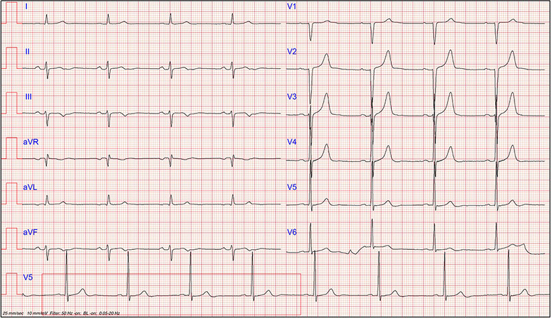
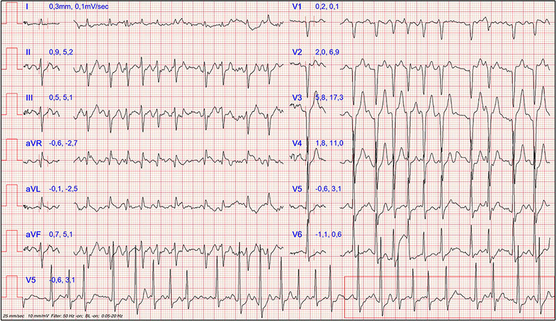
Quiz-18-Fig-1.jpg

Figure 2:

Peak stress ECG: the subject underwent incremental cycle ergometer testing. He reached the maximum load at 285 watts and presented this peak ECG.

Quiz-18-Fig-2.jpg

Test your knowledge


Note: The views and opinions expressed on this page are those of the author and may not be accepted by others. While every attempt is made to keep the information up to date, there is always going to be a lag in updating information. The reader is encouraged to read this in conjunction with appropriate ESC Guidelines. The material on this page is for educational purposes and is not for use as a definitive management strategy in the care of patients. Quiz material on the site are only examples and do not guarantee outcomes from formal examinations.

References

1. Drezner JA, Sharma S, Baggish A, Papadakis M, Wilson MG, Prutkin JM, La Gerche A, Ackerman MJ, Borjesson M, Salerno JC, Asif IM, Owens DS, Chung EH, Emery MS, Froelicher VF, Heidbuchel H, Adamuz C, Asplund CA, Cohen G, Harmon KG, Marek JC, Molossi S, Niebauer J, Pelto HF, Perez MV, Riding NR, Saarel T, Schmied CM, Shipon DM, Stein R, Vetter V, Pelliccia A, Corrado D. International criteria for electrocardiographic interpretation in athletes: Consensus statement British Journal of Sports Medicine 2017;51:704-731.

2. Zema MJ, Kligfield P. ECG poor R-wave progression: review and synthesis. Arch Intern Med. 1982 Jun;142(6):1145-8. PMID: 6212033.

3. Pelliccia A, Sharma S, Gati S, Bäck M, Börjesson M, Caselli S, Collet JP, Corrado D, Drezner JA, Halle M, Hansen D, Heidbuchel H, Myers J, Niebauer J, Papadakis M, Piepoli MF, Prescott E, Roos-Hesselink JW, Stuart AG, Taylor RS, Thompson PD, Tiberi M, Vanhees L, Wilhelm M, ESC Scientific Document Group. 2020 ESC Guidelines on sports cardiology and exercise in patients with cardiovascular disease: The Task Force on sports cardiology and exercise in patients with cardiovascular disease of the European Society of Cardiology (ESC), European Heart Journal, Volume 42, Issue 1, 1 January 2021, Pages 17–96, https://doi.org/10.1093/eurheartj/ehaa605.   

4. Corrado D, Drezner JA, D'Ascenzi F, Zorzi A. How to evaluate premature ventricular beats in the athlete: critical review and proposal of a diagnostic algorithm British Journal of Sports Medicine 2020;54:1142-1148.

5. Perazzolo Marra M, Basso C, De Lazzari M, Rizzo S, Cipriani A, Giorgi B, Lacognata C, Rigato I, Migliore F, Pilichou K, Cacciavillani L, Bertaglia E, Frigo AC, Bauce B, Corrado D, Thiene G, Iliceto S. Morphofunctional Abnormalities of Mitral Annulus and Arrhythmic Mitral Valve Prolapse. Circ Cardiovasc Imaging. 2016 Aug;9(8):e005030. doi: 10.1161/CIRCIMAGING.116.005030. PMID: 27516479; PMCID: PMC4991345.

Notes to editor

Author information:

  • Prof. Dalle Carbonare, Luca G. University Hospital of Verona, Director of the Graduate School of Sport Medicine and Exercise, Dpt. Of Medicine - Clinical Network of Sports and Exercise Medicine of the Veneto Region, Padova, Italy
  • Dr. Martini, Davide University Hospital of Verona, School of Sport Medicine and Exercise, Dpt. Of Medicine - Clinical Network of Sports and Exercise Medicine of the Veneto Region, Padova, Italy
  • Dr. Braggio, Michele University Hospital of Verona, School of Sport Medicine and Exercise, Dpt. Of Medicine - Clinical Network of Sports and Exercise Medicine of the Veneto Region, Padova, Italy