In order to bring you the best possible user experience, this site uses Javascript. If you are seeing this message, it is likely that the Javascript option in your browser is disabled. For optimal viewing of this site, please ensure that Javascript is enabled for your browser.
Did you know that your browser is out of date? To get the best experience using our website we recommend that you upgrade to a newer version. Learn more.

Severe aortic valve stenosis in lower-risk patients: from bicuspid aortic valve to uncommon aetiologies

Reflecting the developing literature regarding transcatheter aortic valve implantation (TAVI) in lower-risk populations, the 2017 guidelines have changed so that patients at intermediate and high clinical risk are directed towards the therapy best suited to providing durable and effective long-term relief of obstruction at the lowest risk. A full evaluation of the patient including the surgical, anaesthetic, medical and social risks, as well as a detailed analysis of the imaging data, is required by the Heart Team in order to make its recommendations. This article discusses three sets of circumstances where decision making is more complicated than it might be for degenerative aortic stenosis, and illustrates how various factors influence the final decision on interventional strategy.

Cardiovascular Surgery
Interventional Cardiology and Cardiovascular Surgery

Introduction

Transcutaneous techniques have revolutionised the management of aortic stenosis (AS) [1]. The original PARTNER trial data demonstrated the effectiveness of transcatheter aortic valve implantation (TAVI) in patients without surgical options, and the equivalence of TAVI in high-risk scenarios [2,3]. More recently, the effectiveness of TAVI (particularly delivered by the femoral route) in intermediate-risk patients has been demonstrated [4,5]. This is changing the paradigm of care from “surgery first until the risks become too high” to “optimum strategy to give the best result in the individual patient". This is reflected in the most recent ESC/EACTS guidelines (2017). Both surgery and TAVI have their merits. A better understanding of these allows an individualised decision-making process to get the best long-term outcomes. Amongst patients at high and low risk, the treatment algorithms are clear. This article addresses patients who have AS which is not degenerative or has other associated valvular lesions and who require a more individualised approach.

Bicuspid disease

Contemporary estimates suggest that 0.5% to 2% of the population have a bicuspid aortic valve with a strong male preponderance [6]. It is associated with more rapid development of degenerative lesions (stenosis or regurgitation). Consequently, younger cohorts have a high proportion of bicuspid cases, typically between 40 and 50%. However, bicuspid disease is present in up to 20% of patients over the age of 80 [7]. It can be subtle to diagnose, particularly when the fusion of the two cusps forms a fibrous, or even calcific, raphe (Figure 1). Both echocardiography and computed tomography (CT) can be misleading [8], particularly when the valve is examined in diastole (closed), where it can be difficult to distinguish a raphe from a thickened cusp margin. Examining systolic frames and the orifice shape is strongly recommended [9]. 


15_27_Figure 1.jpg


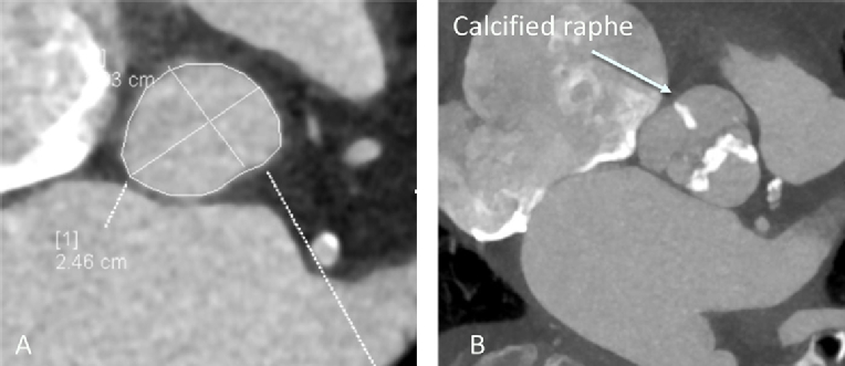
Figure 1.  A) Gated CT demonstrating accurate assessment of annular asymmetry. B) Systolic gated CT frame at the level of the aortic valve demonstrating calcification of the raphe dividing the non-coronary and right coronary cusps. This can easily be misdiagnosed as a tricuspid valve.


The anatomy poses significant potential problems for TAVI. The virtual aortic valve annulus is both large and asymmetric (Figure 1), leading to more complicated valve sizing and the potential risk of regurgitation, which is poorly tolerated in AS [10]. The valve may be less calcified, giving a higher risk of valve displacement. The presence of aortopathy in up to 70% of cases [11,12] is an additional consideration, especially where the aortic dimensions exceed 4.5 cm [13]. Emerging valve design and pre-procedural planning should address these limitations.

Valve sizing

Three-dimensional (3D) techniques are essential for valve sizing. CT is the preferred imaging modality because of its isotropic voxel size and clear tissue interfaces. This allows a simultaneous assessment of valve annulus dimension and eccentricity, leaflet and subvalvular calcification, coronary height, and the size of the ascending aorta. Where CT is either unavailable or contraindicated, 3D transoesophageal echocardiography (TOE) provides a well-validated alternative [14].

Outcome of interventions

Bicuspid valves have been excluded from the randomised clinical trials of TAVI.  Outcome and effectiveness data therefore come either from single site experience or registries, most of which have relatively small numbers. Mortality after TAVI does not seem to be affected by the presence of a bicuspid valve. Only in the Milan experience was late mortality higher, but this was explained by features not related to the valve implant [15]. The risk of aortic regurgitation using first- and second-generation TAVI valves is higher. The exact risk is difficult to define because of a lack of unified definitions. In the TAV-in-BAV registry, the risk of post-procedural significant AR was 28%, although this dropped with CT-based annular sizing [16]. Yoon and colleagues report a much lower risk of significant aortic regurgitation, although this was still higher in the bicuspid patients (10.4% vs. 6.8%). This suggests a difference in definition. This registry confirms a small but significant risk of complications including conversion to surgery and second valve implants. It also confirms that third-generation percutaneous devices have a lower risk of paraprosthetic regurgitation [17]. While the effectiveness of surgical valve replacement is not in doubt, surgical advances such as the sutureless valve appear to be equally effective in bicuspid disease [18].

Take home messages

  • In the absence of randomised evidence, surgical replacement should be the standard of care for patients with bicuspid disease, in particular if the ascending aortic dimensions exceed 4.5 cm. 
  • Where patient factors predict a very high surgical complication risk, TAVI with current techniques is an acceptable alternative.
  • TAVI requires careful attention to valve sizing using 3D techniques and the availability of a range of devices to minimise the risk of post-procedural aortic regurgitation and other complications.

Mitral regurgitation

Patients with AS frequently have coexistent mitral regurgitation (MR). In the PARTNER series of studies, this was observed in more than 20% of patients [19]. This can be due to primary disease of the mitral valve (PMR) or secondary restriction due to changes in annular shape or ventricular geometry (functional mitral regurgitation [FMR]). Increasingly, both primary and secondary elements are present, which makes true dichotomisation challenging. 

Pathophysiology

Changes in ventricular geometry, systolic atrioventricular pressure gradients, and the mitral annular relations mean that the effect of the relief of AS is unpredictable. While initial reports from the PARTNER study suggested that patients undergoing TAVI did better than those undergoing surgical valve replacement [19], most studies have demonstrated that the presence of MR predicts an adverse outcome compared with those who have only minor or no regurgitation. In a meta-analysis of 13 studies, Sanino et al [20] demonstrated a mortality deficit at 30 days and at both one and two years, although the study did demonstrate an improvement in MR. Similar findings were observed in the meta-analysis of Chakravarty et al [21], where the overall excess risk attributable to MR at 30 days was 35%. Interestingly, there was no difference between secondary and primary MR in this study. In a single study which opposes these findings, Vollenbroich et al demonstrated a significantly higher mortality amongst those with FMR [22] as well as other markers suggesting adverse remodelling and function such as lower mean transvalvular gradients. Nombela-Franco et al [23] demonstrated that a large proportion of patients with FMR undergoing surgical aortic valve replacement demonstrate an improvement when MR was at least moderate. Similarly, patients with moderate or severe MR undergoing TAVI showed improvement in up to 50% (in those with moderate or more MR), although the results for all grades of MR were more ambiguous.

Assessment of mitral regurgitation

Given the complex nature of both primary and secondary MR in this context, adequate assessment before a proposed procedure is essential. This should be carried out using TOE with the availability of 3D imaging. TOE allows a detailed assessment of valve morphology, in particular subtle degenerative changes that may not be observed during TTE.  In FMR, an assessment of valve tenting, leaflet restriction and annular dimensions may predict an adverse response to surgical annuloplasty. The severity of MR during TOE is only useful if severe, especially in FMR, as changes in loading conditions may lead to erroneous underestimates of MR severity. TOE also allows an assessment of the suitability for percutaneous mitral valve edge-to-edge repair in the event of ongoing clinically relevant regurgitation for those in whom further surgery is not feasible [24].

Take home messages

  • In patients with PMR which is significant, and in whom surgical risks are not preclusive, surgical aortic valve replacement with either repair or replacement of the mitral valve is the optimum strategy. 
  • For secondary mitral regurgitation, the relative contribution of anatomical vs. physiological drivers (such as marked ventricular remodelling or severe valve tenting) should be taken into consideration and quantitative assessment of mitral regurgitation severity should be undertaken.
  • In severe secondary MR, improvement is seen in 50% of cases and no change in the remainder.
  • TAVI or SAVR in isolation remains a reasonable option in FMR as there is no randomised evidence of benefit from the surgical repair/replacement of bystander MR.
  • In inoperable patients, an assessment for the suitability of percutaneous edge-to-edge valve repair should be undertaken if TAVI fails to bring about a successful clinical result.

Radiation-induced aortic valve disease

The widespread use of radiation to treat both solid tumours and haematological malignancy in the thorax results in significant toxicity. While radiation sparing techniques are now widespread, the results of radiation exposure are delayed, by up to twenty years [25]. Those presenting today may have had highly significant exposure in the late 20th century. Fibrotic toxicity affects all structures within the beam including not only the aortic and the mitral valves, but also the myocardium, pericardium, and aorta, as well as causing severe aortic calcification and pulmonary fibrosis. This means that the prognosis and risk associated with patients having cardiac surgery is significantly higher than in equivalent patients without radiation exposure [26]. 

Patient evaluation can be advanced before the role of radiation is recognised.  Echocardiographic features are very typical: in particular, heavy thickening of the aortic/mitral continuity and anterior mitral leaflet should alert the assessor to the possibility of radiotherapy (Figure 2).

15_27_Figure 2.jpg

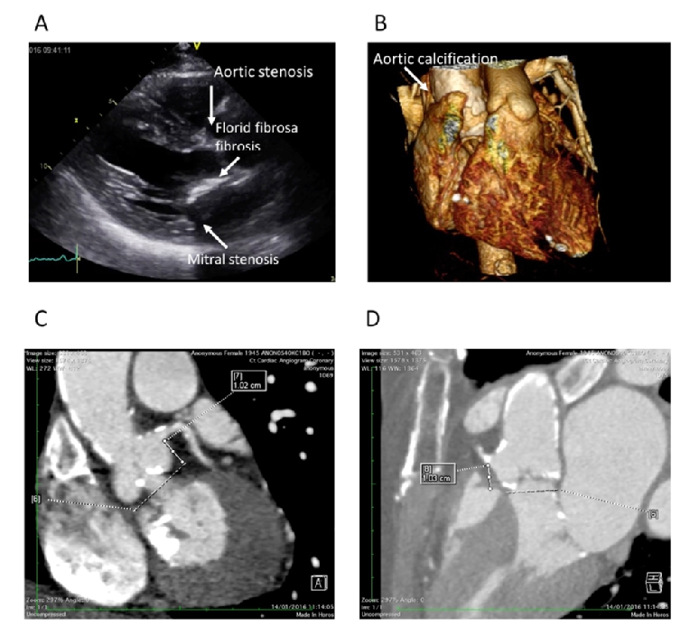
Figure 2.  A) Parasternal long-axis view demonstrating the typical features of radiation-induced heart valve disease. B) 3D volume rendering demonstrating severe circumferential aortic calcification. C) & D) Extensive valve and proximal coronary artery calcification and valvular calcification.


Assessment of radiation-induced heart disease

Evaluation of patients is more detailed than in those with conventional aortic valve disease. A multimodality approach is always required. Assessment of the severity of the aortic valve stenosis is best undertaken using echocardiography. The standard measures of severity apply. However, it is common for patients to express either low flow, or paradoxical low flow pattern (with a transaortic flow rate of less than 35 mls/m2) because of radiotherapy-induced myocardial dysfunction. When the ejection fraction is preserved, significant LV dysfunction can still be demonstrated by careful attention to longitudinal function, in particular global longitudinal strain (27).

Gated CT evaluation of the chest is mandatory. This offers an opportunity to confirm the severity of the aortic valve stenosis, although using the aortic valve Agatston score is not validated in this context. The proximal coronary arteries can be examined, although the high level of calcification at and around the aortic root may make accurate assessment of the coronary difficult, signalling the need for invasive angiography. The CT assessment should include the thoracic aorta to describe the extent and location of aortic calcification and how this may affect surgical access. Finally, using the appropriate lung windows an assessment of the location and extent of pulmonary fibrosis should be undertaken.

In contradistinction to their high surgical mortality, patients with radiation-induced aortic valve disease appear to do as well as those with degenerative aortic stenosis after TAVI [28]. Mitral stenosis is also more common and observed in over 50% of cases but, despite this, the functional improvement is equivalent [29].

Take home messages

  • Patients with chest radiation exposure have a significant delayed risk of AS.
  • Pathology is rarely confined to the aortic valve; the mitral valve is frequently involved, as are the proximal coronary arteries and the ascending aorta. 
  • Surgery is at considerably higher risk.
  • Assessment should be made in a centre with considerable expertise. The availability of gated CT and both 2D and 3D echocardiography is essential.
  • TAVI is associated with a high level of technical and clinical success and is the preferred treatment strategy, assuming mitral valve disease does not dominate the clinical presentation; the risk of post-procedural mitral stenosis remains higher.

Conclusions

AS remains a common disease which requires timely and appropriate therapy. The emerging evidence base supporting the long-term clinical efficacy and durability of TAVI has resulted in a philosophical change, highlighted in the 2017 ESC/EACTS guidelines. These shift the emphasis from age alone in favour of selecting the most appropriate technique for the best long-term results mediated by the multidisciplinary Heart Team. The guidance points towards certain factors that should sway clinical decision making. The circumstances outlined in this article are cases in point and all highlight the need for meticulous diagnostic work-up. Integrating detailed echocardiography and the cross-sectional imaging from CT will assist the Heart Team to make the most appropriate management recommendations.


References


  1. Cribier A, Eltchaninoff H, Tron C, Bauer F, Agatiello C, Sebagh L, Bash A, Nusimovici D, Litzler PY, Bessou JP, Leon MB. Early experience with percutaneous transcatheter implantation of heart valve prosthesis for the treatment of end-stage inoperable patients with calcific aortic stenosis. J Am Coll Cardiol. 2004 Feb 18;43(4):698-703. 
  2. Leon MB, Smith CR, Mack M, Miller DC, Moses JW, Svensson LG, Tuzcu EM, Webb JG, Fontana GP, Makkar RR, Brown DL, Block PC, Guyton RA, Pichard AD, Bavaria JE, Herrmann HC, Douglas PS, Petersen JL, Akin JJ, Anderson WN, Wang D, Pocock S; PARTNER Trial Investigators. Transcatheter aortic-valve implantation for aortic stenosis in patients who cannot undergo surgery. N Engl J Med. 2010 Oct 21;363(17):1597-607. 32
  3. Smith CR, Leon MB, Mack MJ, Miller DC, Moses JW, Svensson LG, Tuzcu EM, Webb JG, Fontana GP, Makkar RR, Williams M, Dewey T, Kapadia S, Babaliaros V, Thourani VH, Corso P, Pichard AD, Bavaria JE, Herrmann HC, Akin JJ, Anderson WN, Wang D, Pocock SJ; PARTNER Trial Investigators. Transcatheter versus surgical aortic-valve replacement in high-risk patients. N Engl J Med. 2011 Jun 9;364 (23):2187-98.
  4. Leon MB, Smith CR, Mack MJ, Makkar RR, Svensson LG, Kodali SK, Thourani VH, Tuzcu EM, Miller DC, Herrmann HC, Doshi D, Cohen DJ, Pichard AD, Kapadia S, Dewey T, Babaliaros V, Szeto WY, Williams MR, Kereiakes D, Zajarias A, Greason KL, Whisenant BK, Hodson RW, Moses JW, Trento A, Brown DL, Fearon WF, Pibarot P, Hahn RT, Jaber WA, Anderson WN, Alu MC, Webb JG; PARTNER 2 Investigators. Transcatheter or Surgical Aortic-Valve Replacement in Intermediate- Risk Patients. N Engl J Med. 2016 Apr 28;374(17):1609-20.
  5. Auffret V, Lefevre T, Van Belle E, Eltchaninoff H, Iung B, Koning R, Motreff P, Leprince P, Verhoye JP, Manigold T, Souteyrand G, Boulmier D, Joly P, Pinaud F, Himbert D, Collet JP, Rioufol G, Ghostine S, Bar O, Dibie A, Champagnac D, Leroux L, Collet F, Teiger E, Darremont O, Folliguet T, Leclercq F, Lhermusier T, Olhmann P, Huret B, Lorgis L, Drogoul L, Bertrand B, Spaulding C, Quilliet L, Cuisset T, Delomez M, Beygui F, Claudel JP, Hepp A, Jegou A, Gommeaux A, Mirode A, Christiaens L, Christophe C, Cassat C, Metz D, Mangin L, Isaaz K, Jacquemin L, Guyon P, Pouillot C, Makowski S, Bataille V, Rodés-Cabau J, Gilard M, Le Breton H; FRANCE TAVI Investigators. Temporal Trends in Transcatheter Aortic Valve Replacement in France: FRANCE 2 to FRANCE TAVI. J Am Coll Cardiol. 2017 Jul 4;70(1):42-55.
  6. Basso C, Boschello M, Perrone C, Mecenero A, Cera A, Bicego D, Thiene G, De Dominicis E. An echocardiographic survey of primary school children for bicuspid aortic valve. Am J Cardiol. 2004 Mar 1;93(5):661-3.
  7. Roberts WC, Janning KG, Ko JM, Filardo G, Matter GJ. Frequency of congenitally bicuspid aortic valves in patients ≥80 years of age undergoing aortic valve replacement for aortic stenosis (with or without aortic regurgitation) and implications for transcatheter aortic valve implantation. Am J Cardiol. 2012 Jun 1;109(11):1632-6.
  8. Hillebrand M, Koschyk D, Ter Hark P, Schüler H, Rybczynski M, Berger J, Gulati A, Bernhardt AM, Detter C, Girdauskas E, Blankenberg S, von Kodolitsch Y. Diagnostic accuracy study of routine echocardiography for bicuspid aortic valve: a retrospective study and meta-analysis. Cardiovasc Diagn Ther. 2017 Aug;7(4):367-379.
  9. Schoenhagen P, Hausleiter J, Achenbach S, Desai MY, Tuzcu EM. Computed tomography in the evaluation for transcatheter aortic valve implantation (TAVI). Cardiovasc Diagn Ther. 2011 Dec 1;1(1):44-56.
  10. Kodali SK, Williams MR, Smith CR, Svensson LG, Webb JG, Makkar RR, Fontana GP, Dewey TM, Thourani VH, Pichard AD, Fischbein M, Szeto WY, Lim S, Greason KL, Teirstein PS, Malaisrie SC, Douglas PS, Hahn RT, Whisenant B, Zajarias A, Wang D, Akin JJ, Anderson WN, Leon MB; PARTNER Trial Investigators. Two-year outcomes after transcatheter or surgical aortic-valve replacement. N Engl J Med. 2012 May 3;366(18):1686-95.
  11. Evangelista A, Gallego P, Calvo-Iglesias F, Bermejo J, Robledo-Carmona J, Sánchez V, Saura D, Arnold R, Carro A, Maldonado G, Sao-Avilés A, Teixidó G, Galian L, Rodríguez-Palomares J, García-Dorado D. Anatomical and clinical predictors of valve dysfunction and aortic dilation in bicuspid aortic valve disease. Heart. 2017 Sep 1. pii: heartjnl-2017-311560. doi: 10.1136/heartjnl-2017-311560. [Epub ahead of print].
  12. Jackson V, Petrini J, Caidahl K, Eriksson MJ, Liska J, Eriksson P, Franco-Cereceda A. Bicuspid aortic valve leaflet morphology in relation to aortic root morphology: a study of 300 patients undergoing open-heart surgery. Eur J Cardiothorac Surg. 2011 Sep;40(3):e118-24.
  13. Vahanian A, Alfieri O, Andreotti F, Antunes MJ, Barón-Esquivias G, Baumgartner H, Borger MA, Carrel TP, De Bonis M, Evangelista A, Falk V, Lung B, Lancellotti P, Pierard L, Price S, Schäfers HJ, Schuler G, Stepinska J, Swedberg K, Takkenberg J, Von Oppell UO, Windecker S, Zamorano JL, Zembala M; ESC Committee for Practice Guidelines (CPG); Joint Task Force on the Management of Valvular Heart Disease of the European Society of Cardiology (ESC); European Association for Cardio-Thoracic Surgery (EACTS).  Guidelines on the management of valvular heart disease (version 2012): the Joint Task Force on the Management of Valvular Heart Disease of the European Society of Cardiology (ESC) and the European Association for Cardio-Thoracic Surgery (EACTS). Eur J Cardiothorac Surg. 2012 Oct;42(4):S1-44.
  14. Bleakley C, Eskandari M, Monaghan M. 3D transoesophageal echocardiography in the TAVI sizing arena: should we do it and how do we do it? Echo Res Pract. 2017 Mar;4(1):R21-R32.
  15. Costopoulos C, Latib A, Maisano F, Testa L, Bedogni F, Buchanan L, Naganuma T, Sticchi A, Sato K, Miyazaki T, Figini F, Giannini F, Taramasso M, Naim C, Carlino M, Chieffo A, Montorfano M, Alfieri O, Colombo A. Comparison of results of transcatheter aortic valve implantation in patients with severely stenotic bicuspid versus tricuspid or nonbicuspid valves. Am J Cardiol. 2014 Apr 15;113(8):1390-3.
  16. Mylotte D, Lefevre T, Søndergaard L, Watanabe Y, Modine T, Dvir D, Bosmans J, Tchetche D, Kornowski R, Sinning JM, Thériault-Lauzier P, O'Sullivan CJ, Barbanti M, Debry N, Buithieu J, Codner P, Dorfmeister M, Martucci G, Nickenig G, Wenaweser P, Tamburino C, Grube E, Webb JG, Windecker S, Lange R, Piazza N. Transcatheter aortic valve replacement in bicuspid aortic valve disease. J Am Coll Cardiol. 2014 Dec 9;64(22):2330-9.
  17. Yoon SH, Bleiziffer S, De Backer O, Delgado V, Arai T, Ziegelmueller J, Barbanti M, Sharma R, Perlman GY, Khalique OK, Holy EW, Saraf S, Deuschl F, Fujita B, Ruile P, Neumann FJ, Pache G, Takahashi M, Kaneko H, Schmidt T, Ohno Y, Schofer N, Kong WKF, Tay E, Sugiyama D, Kawamori H, Maeno Y, Abramowitz Y, Chakravarty T, Nakamura M, Kuwata S, Yong G, Kao HL, Lee M, Kim HS, Modine T, Wong SC, Bedgoni F, Testa L, Teiger E, Butter C, Ensminger SM, Schaefer U, Dvir D, Blanke P, Leipsic J, Nietlispach F, Abdel-Wahab M, Chevalier B, Tamburino C, Hildick-Smith D, Whisenant BK, Park SJ, Colombo A, Latib A, Kodali SK, Bax JJ, Søndergaard L, Webb JG, Lefèvre T, Leon MB, Makkar R. Outcomes in Transcatheter Aortic Valve Replacement for Bicuspid Versus Tricuspid Aortic Valve Stenosis. J Am Coll Cardiol. 2017 May 30;69(21):2579-2589.
  18. Nguyen A, Fortin W, Mazine A, Bouchard D, Carrier M, El Hamamsy I, Lamarche Y, Demers P. Sutureless aortic valve replacement in patients who have bicuspid aortic valve. J Thorac Cardiovasc Surg. 2015 Oct;150(4):851-7.
  19. Barbanti M, Webb JG, Hahn RT, Feldman T, Boone RH, Smith CR, Kodali S, Zajarias A, Thompson CR, Green P, Babaliaros V, Makkar RR, Szeto WY, Douglas PS, McAndrew T, Hueter I, Miller DC, Leon MB; Placement of Aortic Transcatheter Valve Trial Investigators. Impact of preoperative moderate/severe mitral regurgitation on 2-year outcome after transcatheter and surgical aortic valve replacement: insight from the Placement of Aortic Transcatheter Valve (PARTNER) Trial Cohort A. Circulation. 2013 Dec 24;128(25):2776-84.
  20. Sannino A, Losi MA, Schiattarella GG, Gargiulo G, Perrino C, Stabile E, Toscano E, Giugliano G, Brevetti L, Franzone A, Cirillo P, Imbriaco M, Trimarco B, Esposito G. Meta-analysis of mortality outcomes and mitral regurgitation evolution in 4,839 patients having transcatheter aortic valve implantation for severe aortic stenosis. Am J Cardiol. 2014 Sep 15;114(6):875-82.
  21. Chakravarty T, Van Belle E, Jilaihawi H, Noheria A, Testa L, Bedogni F, Rück A, Barbanti M, Toggweiler S, Thomas M, Khawaja MZ, Hutter A, Abramowitz Y, Siegel RJ, Cheng W, Webb J, Leon MB, Makkar RR. Meta-analysis of the impact of mitral regurgitation on outcomes after transcatheter aortic valve implantation. Am J Cardiol. 2015 Apr 1;115(7):942-9.
  22. Vollenbroich R, Stortecky S, Praz F, Lanz J, Franzone A, Zuk K, Heg D, Valgimigli M, O'Sullivan CJ, Heinisch C, Roost E, Wenaweser P, Windecker S, Pilgrim T. The impact of functional vs degenerative mitral regurgitation on clinical outcomes among patients undergoing transcatheter aortic valve implantation. Am Heart J. 2017 Feb; 184:71-80.
  23. Nombela-Franco L, Ribeiro HB, Urena M, Allende R, Amat-Santos I, DeLarochellière R, Dumont E, Doyle D, DeLarochellière H, Laflamme J, Laflamme L, García E, Macaya C, Jiménez-Quevedo P, Côté M, Bergeron S, Beaudoin J, Pibarot P, Rodés-Cabau J. Significant mitral regurgitation left untreated at the time of aortic valve replacement: a comprehensive review of a frequent entity in the transcatheter aortic valve replacement era. J Am Coll Cardiol. 2014 Jun 24;63(24):2643-58.
  24. Braun D, Frerker C, Körber MI, Gaemperli O, Patzelt J, Schaefer U, Hammerstingl C, Boekstegers P, Ott I, Ince H, Thiele H, Hausleiter J. Percutaneous Edge-to-Edge Repair of Recurrent Severe Mitral Regurgitation After Surgical Mitral Valve Repair. J Am Coll Cardiol. 2017 Jul 25;70(4):504-505.
  25. Gujral DM, Lloyd G, Bhattacharyya S. Radiation-induced valvular heart disease. Heart. 2016 Feb 15;102(4):269-76. g
  26. Wu W, Masri A, Popovic ZB, Smedira NG, Lytle BW, Marwick TH, Griffin BP, Desai MY. Long-term survival of patients with radiation heart disease undergoing cardiac surgery: a cohort study. Circulation. 2013 Apr 9;127(14):1476-85.
  27. Lancellotti P, Nkomo VT, Badano LP, Bergler-Klein J, Bogaert J, Davin L, Cosyns B, Coucke P, Dulgheru R, Edvardsen T, Gaemperli O, Galderisi M, Griffin B, Heidenreich PA, Nieman K, Plana JC, Port SC, Scherrer-Crosbie M, Schwartz RG, Sebag IA, Voigt JU, Wann S, Yang PC; European Society of Cardiology Working Groups on Nuclear Cardiology and Cardiac Computed Tomography and Cardiovascular Magnetic Resonance; American Society of Nuclear Cardiology; Society for Cardiovascular Magnetic Resonance; Society of Cardiovascular Computed Tomography. Expert consensus for multi-modality imaging evaluation of cardiovascular complications of radiotherapy in adults: a report from the European Association of Cardiovascular Imaging and the American Society of Echocardiography. Eur Heart J Cardiovasc Imaging. 2013 Aug;14(8):721-40.
  28. Bouleti C, Amsallem M, Touati A, Himbert D, Iung B, Alos B, Brochet E, Urena M, Ghodbane W, Ou P, Dilly MP, Nataf P, Vahanian A. Early and late outcomes after trans-catheter aortic valve implantation in patients with previous chest radiation. Heart. 2016 Jul 1;102(13):1044-51.
  29. Dijos M, Reynaud A, Leroux L, Réant P, Cornolle C, Roudaut R, Dos Santos P, Lafitte S. Efficacy and follow-up of transcatheter aortic valve implantation in patients with radiation-induced aortic stenosis. Open Heart. 2015 Aug 25;2(1): e000252.

Notes to editor


Authors:

Guy Lloyd1, MD, FRCP; David Messika-Zeitoun2, MD, PhD

  1. Barts Heart Centre, St. Bartholomew’s Hospital, West Smithfield, London, United Kingdom;
  2. Bichat Hospital and University Paris VII, Paris, France


Author for correspondence:

Dr. Guy Lloyd, MD, FRCP
Barts Heart Centre, St Bartholomew's Hospital, West Smithfield, London EC1A 7BA, United Kingdom

E-mail: glloyd@nhs.net


Author disclosures:

Dr. Guy Lloyd - speaking engagements and advisory boards - Edwards, Philips, GE; research grants - Medtronic. Professor David Messika-Zeitoun - consultant for Edwards, Valtech, Mardil and Cardiawave; research grants from Edwards and Abbott.


The content of this article reflects the personal opinion of the author/s and is not necessarily the official position of the European Society of Cardiology.