In order to bring you the best possible user experience, this site uses Javascript. If you are seeing this message, it is likely that the Javascript option in your browser is disabled. For optimal viewing of this site, please ensure that Javascript is enabled for your browser.
Did you know that your browser is out of date? To get the best experience using our website we recommend that you upgrade to a newer version. Learn more.

Bicuspid aortic valve

An article from the e-Journal of Cardiology Practice

Bicuspid aortic valve may be complicated with aortic valve stenosis and/or regurgitation in addition to risk of endocarditis. Furthermore, it is associated with coarctation and aortopathy that may result in aortic aneurysm, dissection or rupture.
The definitive treatment is surgery for the valve and/or aortic root depending on severity of valve lesion, aortic diameter and rate of progression of aortopathy. 

Valvular Heart Disease

Background

Bicuspid aortic valve (BAV) is the most common congenital cardiac abnormality, affecting approximately 1%-2% of the general population. (1,2) Adverse cardiac outcomes related to the valve and/or root (3) put a large burden on cardiovascular patients, in addition to the many undergoing aortic valve replacement who will also need aortic root surgery. 
Although inheritance is variable, it is a familial disease (4,5) with an estimated 10% chance of a first degree relative being affected (6,7) and 20%-30% when aortopathy is included into the calculation. (8,9) Additionally, certain gene mutations (NOTCH1) have been associated. 
The BAV is composed of not three aortic normal leaflets - as in the normal tricuspid valve - but two abnormal leaflets, usually unequal in size, possibly resulting from inadequate fibrillin-1 production during valvulogenesis. (10)
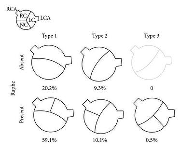
The most common form has the two commissures located in an anteroposterior direction giving left and right cusps. Less common is having the commissures located on the right and left sides of the annulus leading to anterior and posterior cusps. Type 1 BAVs are more likely to stenose as adults while type 2 valves will have complications earlier. Rarely, there is fusion of the left and non-coronary cusps. A raphe is present on the right and anterior cusps, respectively, and this can make the valve appear tricuspid on echocardiography (Figure 1). (11)

Figure 1: Classification and Incidence of BAV.


Complications

Complications are 1) aortic stenosis 2) aortic regurgitation (or incompetence) 3) endocarditis. Calcification usually also occurs.
Due to abnormal stresses, even a normally functioning BAV can progress and damage gradually to reveal abnormal folding and creasing throughout the cardiac cycle with more turbulent flow, as well as restricted motion. (12)
Aortopathy and coarctation have implications on the prognosis of BAV. In several cohort studies, the long-term survival of patients was similar to the normal population, but with more cardiac events and need for surgical interventions on the valve and/or aorta. (13,14) Natural history of BAV ranges from severe aortic stenosis in childhood to longstanding asymptomatic disease into the elderly years.

  • Aortic stenosis is the most common complication and may require surgery, most often necessary in  middle age. (15,16) Only 15% of patients with BAV will have a normal function in the fifth decade.
  • Aortic regurgitation also occurs due to the abnormality and may present with or without aortic stenosis. (17) Various aetiologies have been suggested to explain the mechanism of aortic regurgitation in BAV, including cusp prolapse, endocarditis, dilated aortic root, and myxoid degeneration of the valve. (18)
  • Endocarditis is more common in BAV than in the normal tricuspid aortic valve. The incidence is estimated to be less than 2% in different series, but the outcome of endocarditis tends to be worse than in normal valves.


Aortopathy

Aortopathy refers to 'dilated aortic root'. It is usually associated with BAV and may lead to aortic aneurysm, dissection and rupture. (19,20) The prevalence of ascending aorta dilatation in patients with BAV ranged from 7.5% to 79%, according to the study of aortas in the population and region examined. (21-22) Incidence was higher in patients with fusion of the right and non-coronary cusps than the more common phenotype of fusion of the right and left coronary cusps. The mechanism of aortopathy is explained by genetic theory (aortic root fragility and elastic fragmentation due to inherited developmental defect) and hemodynamic theory (abnormal stress on aortic root due to turbulent flow through BAV). (23-27)
Three different shapes of proximal aorta in BAV were described and co-related to the type of BAV cusp configuration. (28) Serial echocardiographic examinations found higher rates of growth at all levels of the proximal aorta above the annulus in patients with BAV in comparison to those with tricuspid aortic valves. (29-31) Aortic dissection is a lethal sequel in patients with BAV and occurs at a younger age. Recent data suggest that BAV increases the risk of aortic dissection nine-fold over that in the general population. (32).Its incidence is less than 1% more in some studies, yet reaches 4% in others. (33)
It is worth noting that aortic root dilatation is an important predictor of dissection and rupture in BAV-associated aneurysms, even if the aortic valve is functioning normally. (34)

Coarctation of aorta

Coarctation of aorta is another major abnormality that can be associated with 20% or more of BAV. In the presurgical era, this association was found to increase the risk of death from aortic dissection more than if coarctation existed without BAV. (35) Coarctation is sometimes regarded as a part of the diffuse aortopathy associated with BAV.
Less common congenital anomalies associated with BAV include ventricular septal defects, Ebstein’s anomaly, hypoplastic left heart syndrome, abnormal coronary anatomy, patent ductus arteriosus, atrial septal defects and bicuspid pulmonary valve. 

Diagnosis

Echocardiography: The mainstay of diagnosis is echocardiography (transthoracic or transoesophageal) which can provide a definitive diagnosis in the majority of patients with 92% sensitivity and 96% specificity when images are adequate (36). The echocardiographic report should adequately evaluate aortic valve morphology and measure the severity of any stenosis or regurge. It should also include aortic measurements at the aortic annulus, sinuses, sinotubular junction, and mid-ascending aorta.
Doppler: In addition, evaluation for aortic coarctation using Doppler interrogation of the proximal descending aorta should be performed. Aortic magnetic resonance angiography or CT angiography is indicated if morphology of the aortic root and/or ascending aorta cannot be assessed accurately or fully by echocardiography.
As recommended by the recent north American and European guidelines, patients with BAV who have documented dilation of aorta should have serial assessment of aortic morphology to detect progression of aortopathy early enough before complications occur (37). Recent tools for diagnosis and risk stratification have been suggested using metalloproteinase plasma assays and magnetic resonance imaging (38).

Treatment

Aortic valve replacement is the only definitive treatment for BAV associated with severe stenosis and/or regurgitation. The indications for surgery are similar to those with tricuspid aortic valve.
New techniques of repair such as transcatheter aortic valve implantation have also been reported in BAV (39) but are still considered as a relative contraindication in recent guidelines. In patients who refuse surgery or are not candidates for surgical intervention, medical therapy is limited and is mainly used to treat existent cardiac conditions such as heart failure, coronary artery disease or hypertension.
However, there is a possible role of beta blockers and ARBs in delaying progression of aortic dilatation, extrapolating from patients with aortopathy associated with Marfan’s syndrome, but have not been confirmed in clinical studies. 

Guidelines 

The recent ESC guidelines for valvular heart disease management recommend that sugery should be considered for any patient with aortic root diameter of ≥55 mm (Table 1). However, in patients with BAV, surgery in aortic  diameters of ≥50 mm should be based on patient age, body size, comorbidities, type of surgery, and the presence of additional risk factors  - family history, systemic hypertension, coarctation of the aorta, or increase in aorticdiameter ≥2 mm/year in repeated examinations using the same technique and confirmed by another technique.
The new ACC/AHA guidelines recommend surgical repair of aortic root if aortic root diameter is ≥5.5 cm (Class I). This recommendation changed in 2014 from the previous 2006 recommendation which had a cutoff value of 5 cm.
It should also be noted that the ACC/AHA guidelines considered the evidence of rapid progression of a maximum aortic cross-section: body/height ratio of 10 cm2/m was proposed as an additional measurement to decide for aortic root surgery and was found to be associated with an increased risk for aortic dissection in several studies. (40-42)

Table 1. Indications of Aortic Root Surgery in Patients with BAV according to Measured Aortic Root Diameter

Aortic Root Diameter Indication for Elective Aortic Root Surgery
≥55 mm  All - class I in ACC/AHA guidelines and IIa in ESC guidelines
50-54 mm  If associated with any of:
  • Family history of dissection or unexplained sudden death
  • Rapid growth progression of aortic dilatation
  • Others (systemic hypertension, associated coarctation, female patient seeking pregnancy)
≥45 mm If concomitant valve surgery is indicated

Conclusions

Bicuspid aortic valve may be complicated with aortic valve stenosis and/or regurgitation, in addition to risk of endocarditis. Furthermore, it is associated with coarctation and aortopathy that may result in aortic aneurysm, dissection or rupture. The definitive treatment is surgery for the valve and/or aortic root, depending on severity of valve lesion, aortic diameter and rate of progression of aortopathy. 

References


1 - Bicuspid aortic valve.
Williams D. S. J Insur Med, 2006;. 38 (1): 72-4
2 - Clinical significance of the bicuspid aortic valve.
Ward C. Heart, 2000. 83 (1): 81-5
3 - Outcomes in adults with bicuspid aortic valves.
Tzemos N., Therrien J., Yip J., Thanassoulis G., Tremblay S., Jamorski M. T., & Webb G. D. JAMA, 2008. 300 (11): 1317–25.
4 - Mutations in NOTCH1 cause aortic valve disease.
Garg V., Muth A. N., Ransom J. F., Schluterman M. K., Barnes R., King I. N., Grossfeld P. D., & Srivastava D. Nature, 2005. 437 (7056): 270–4.
5 - Genetic insights into bicuspid aortic valve formation.
Laforest B., & Nemer M. Cardiol Res Pract, 2012: 180297. doi: 10.1155/2012/180297
6 - Bicuspid aortic valve disease: A comprehensive review.
Mordi I. & Tzemos N. Cardiol Res Pract, 2012: 196037. doi: 10.1155/2012/196037
7 - Bicuspid aortic valve is heritable.
Cripe L., Andelfinger G., Martin L. J., Shooner K., & Benson D. W. J Am Coll Cardiol, 2004, July 7. 44 (1): 138-43.8. 8 - Aortopathy is Prevalent in Relatives of Bicuspid Aortic Valve Patients.  
Biner S., Rafique A. R., Ray I., Cuk O., Siegel R. J., & Tolstrup K. J Am Coll Cardiol, 2009.  53 (24): 2288–95.
9 - 2014 AHA/ACC Guideline for the Management of Patients with Valvular Heart Disease:
A Report of the American College of Cardiology/American Heart Association Task Force on Practice Guidelines. Nishimura R. A, Otto C. M., Bonow R. O., Carabello B. A., Erwin J. P. 3rd, Guyton R. A., O'Gara P. T., Ruiz C. E., Skubas N. J., Sorajja P., Sundt T. M. 3rd, Thomas J. D., Anderson J. L., Halperin J. L., Albert N. M., Bozkurt B., Brindis R. G., Creager M. A.,Curtis L. H., DeMets D., Guyton R. A., Hochman J. S., Kovacs R. J., Ohman E. M., Pressler S. J., Sellke F. W., Shen W. K.,Stevenson W. G., & Yancy C. W.  J Thorac Cardiovasc Surg, 2014, July. 148 (1): e1-e132.
10 - Clinical and pathophysiological implications of a bicuspid aortic valve.
Fedak P. W., Verma S., David T. E., Leask R. L., Weisel R. D., & Butany J. Circulation, 2002. 106 (8): 900-4.
11 - The bicuspid aortic valve: an integrated phenotypic classification of leaflet morphology and aortic root shap.
Schaefer B. M., Lewin M. B., Stout K. K., Gill E., Prueitt A., Byers P. H., & Otto C. M. Heart, 2008. 94 (12): 1634-38. 
12 - The congenitally bicuspid aortic valve: how does it function? Why does it fail?
Robicsek F., Thubrikar M. J., Cook J. W., & Fowler B. Ann Thorac Surg, 2004. 77: 177-85.
13 - Natural history of asymptomatic patients with normally functioning or minimally dysfunctional bicuspid aortic valve in the community.  
Michelena H. I., Desjardins V. A., Avierinos J. F., Russo A., Nkomo V. T, Sundt T. M., Pellikka P. A., Tajik A. J., & Enriquez-Sarano M. Circulation, 2008. 117: 2776-84.
14 - Incidence of aortic complications in patients with bicuspid aortic valves.
Michelena H. I., Khanna A. D., Mahoney D., Margaryan E., Topilsky Y., Suri R. M., Eidem B., Edwards W. D., Sundt T. M. 3rd, & Enriquez-Sarano M. JAMA, 2011. 306 (10): 1104-12.
15 - Left Ventricular Outflow Obstruction: Subaortic Stenosis, Bicuspid Aortic Valve, Supravalvar Aortic Stenosis, and Coarctation of the Aorta.
Aboulhosn J., & Child J. S. Circulation, 2006. 114: 2412–22.
16 - Frequency by decades of unicuspid, bicuspid, and tricuspid aortic valves in adults having isolated aortic valve replacement for aortic stenosis, with or without associated aortic regurgitation.
Roberts W. C. & Ko J. M. Circulation, 2005;. 11: 920–5.
17 - Bicuspid aortic valves are associated with aortic dilatation out of proportion to coexistent valvular lesions.
Keane M. G., Wiegers S. E., Plappert T., Pochettino A., Bavaria J. E., & Sutton M. G. Circulation, 2000. 102: III35-III39. 
18 - Congenital bicuspid aortic valve: analysis of 63 surgical cases.
Yotsumoto G., Moriyama Y., Toyohira H., Shimokawa S., Iguro Y., Watanabe S., Masuda H., Hisatomi K., & Taira A. J. Heart Valve Dis, 1998. 7 (5): 500-3. 
19 - Aortic root dilatation in young men with normally functioning bicuspid aortic valves.
Nistri S., Sorbo M. D., Marin M., Palisi M., Scognamiglio R., & Thiene G. Heart, 1999. 82 (1): 19-22.
20 - Ascending aortic dilatation associated with bicuspid aortic valve: pathophysiology, molecular biology, and clinical implications.
Tadros T. M., Klein M. D., & Shapira O. M. Circulation, 2009. 119 (6): 880-90.
21 - Frequency of aortic root dilation in children with a bicuspid aortic valve. Gurvitz M., Chang R. K., Drant S., & Allada V. Am J Cardiol, 2004. 94: 1337–1340.
22 - Changes in size of ascending aorta and aortic valve function with time in patients with congenitally bicuspid aortic valves.
Ferencik M., & Pape L. A. Am J Cardiol, 2003. 92: 43– 46. 
23 - Bicuspid aortic valve disease: the role of oxidative stress in Lrp5 bone formation.
Rajamannan N. M. Cardiovasc Pathol, 2011. 20 (3): 168-76.
24 - Is aortopathy in bicuspid aortic valve disease a congenital defect or a result of abnormal hemodynamics? A critical reappraisal of a one-sided argument.
Girdauskas E., Borger M. A., Secknus M. A., Girdauskas G., & Kuntze T. Eur J Cardiothorac Surg, 2011. 39 (6): 809-14.
25 - Relation of bicuspid aortic valve morphology to the dilatation pattern of the proximal aorta: focus on the transvalvular flow.
Girdauskas E., Disha K., Borger M. A., & Kuntze T. Cardiol Res Pract, 2012:478259.  doi: 10.1155/2012/478259.
26 - Pathophysiological implications of different bicuspid aortic valve configurations.
Kari F. A., Beyersdorf F., & Siepe M. Cardiol Res Pract, 2012: 735829. doi: 10.1155/2012/735829. 
27 - Increased ascending aortic wall stress in patients with bicuspid aortic valves.
Nathan D. P., Xu C., Plappert T., Desjardins B., Gorman J. H. 3rd, Bavaria J. E., Gorman R. C., Chandran K. B., & Jackson B. M. Ann Thorac Surg, 2011. 92 (4): 1384-9.
28 -  Bicuspid aortic valve disease.
Siu S. C. & Silversides C. K. J Am Coll Cardiol, 2010. 55 (25): 2789-800. 
29 - The bicuspid aortic valve and its relation to aortic dilation.
Yuan S. M, Jing H., & Lavee J. Clinics (Sao Paulo). 2010. 65 (5):497-505.
30 - Failure to prevent progressive dilation of ascending aorta by aortic valve replacement in patients with bicuspid aortic valve: comparison with tricuspid aortic valve.
Yasuda H. I, Nakatani S., Stugaard M., Tsujita-Kuroda Y., Bando K., Kobayashi J., Yamagishi M., Kitakaze M., Kitamura S., & Miyatake K. Circulation, 2003. 108: II291–II294. 
31 - Natural history of ascending aortic aneurysms in the setting of an unreplaced bicuspid aortic valve.
Davies R. R. I., Kaple R. K., Mandapati D., Gallo A., Botta D. M. Jr, Elefteriades J. A., & Coady M.A. Ann Thorac Surg, 2007. 83: 1338–1344. 
32 - Replacement of the Proximal Arch. Should It Be Routine in Patients with Bicuspid Aortic Valve and Ascending Aneurysms
Bolman R. M. III. Tex Heart Inst J, 2011. 38 (6): 672-3.
33 - A critical review of the ACC/AHA practice guidelines on bicuspid aortic valve with dilated ascending aorta.
Guntheroth W. G. Am J Cardiol, 2008. 102: 107-10.
34 - Aortic aneurysm in patients with functionally normal or minimally stenotic bicuspid aortic valve.
Pachulski R. T., Weinberg A. L., & Chan K. L. Am J Cardiol, 1991. 67 (8): 781-2.
35 - Bicuspid aortic valve and coarctation: two villains part of a diffuse problem. Warnes C. A. Heart, 2003. 89 (9): 965-6. 
36 - Diagnostic value of cardiac CT in the evaluation of bicuspid aortic stenosis: comparison with echocardiography and operative findings.
Tanaka R., Yoshioka K., Niinuma H., Ohsawa S., Okabayashi H., & Ehara S. American Journal of Roentgenology, 2010. 195(4): 895–899.
37 - ESC Committee for Practice Guidelines (CPG); Joint Task Force on the Management of Valvular Heart Disease of the European Society of Cardiology (ESC); European Association for Cardio-Thoracic Surgery (EACTS). Guidelines on the management of valvular heart disease (version 2012): the Joint Task Force on the Management of Valvular Heart Disease of the European Society of Cardiology (ESC) and the European Association for Cardio-Thoracic Surgery (EACTS).
Vahanian A., Alfieri O., Andreotti F., Antunes M. J., Barón-Esquivias G., Baumgartner H., Borger M. A., Carrel T. P., De Bonis M., Evangelista A., Falk V., Lung B., Lancellotti P., Pierard L., Price S., Schäfers H. J., Schuler G., Stepinska J., Swedberg K., Takkenberg J., Von Oppell U. O., Windecker S., Zamorano J. L., & Zembala M. Eur J Cardiothorac Surg, 2012. 42 (4): S1-44. doi: 10.1093/ejcts/ezs455
38 - Hemodynamic predictors of aortic dilatation in bicuspid aortic valve by velocity-encoded cardiovascular magnetic resonance.
den Reijer P. M., Sallee D. 3rd, van der Velden P., Zaaijer E. R., Parks W. J., Ramamurthy S., Robbie T. Q., Donati G., Lamphier C., Beekman R. P., & Brummer M.E. J Cardiovasc Magn Reson, 2010. 12:4. doi: 10.1186/1532-429X-12-4 
39 - Transcatheter implantation of an aortic valve prosthesis in a female patient with severe bicuspid aortic stenosis.
Kochman J., Huczek Z., Koltowski L., & Michalak M. Eur Heart J, 2012. 33 1: 112-118.
40 - Current indications for surgical repair in patients with bicuspid aortic valve and ascending aortic ectasia
Etz C. D., Misfeld M., Borger M. A., Luehr M., Strotdrees E., & Mohr F. W. Cardiol Res Pract, 2012: 313879. doi: 10.1155/2012/313879
41 - Bicuspid aortic valve surgery with proactive ascending aorta repair
Svensson L. G. I, Kim K. H, Blackstone E. H., Rajeswaran J., Gillinov A. M., Mihaljevic T., Griffin B. P., Grimm R., Stewart W. J.,Hammer D. F., & Lytle B. W. J Thorac Cardiovasc Surg, 2011. 142 (3): 622-9.
42 - Relationship of aortic cross-sectional area to height ratio and the risk of aortic dissection in patients with bicuspid aortic valves.
Svensson L. G., Kim K. H., Lytle B. W., & Cosgrove D. M. J Thorac Cardiovasc Surg, 2003. 126 (3): 892-3.

Notes to editor


Dr. Adel Shabana, Heart Hospital, Hamad Medical Corporation, Doha, Qatar.

Author's disclosures: None declared.

The content of this article reflects the personal opinion of the author/s and is not necessarily the official position of the European Society of Cardiology.