
Pathophysiology
Orthostatic intolerance syndromes refer to symptoms in which the upright position (most often the movement from sitting or lying to an upright position) causes symptomatic arterial hypotension. Orthostatic intolerance syndromes occur when the autonomic nervous system is incapacitated and fails to respond to the challenges imposed by the upright position.
A second major cause is 'volume depletion' in which the autonomic nervous system is itself not deranged, but is unable to maintain blood pressure due to decreased circulating volume (1).
Drug-induced autonomic failure is probably the most frequent cause of orthostatic intolerance. Diuretics and vasodilators cause central volume depletion; tricyclic antidepressives, phenothiazines, antihistamines, levodopa (Parkinson’s disease) and MAO-inhibitors exert a direct action on central nervous system. The effects of alcohol include both direct acute actions on central nervous system as well as central volume depletion.
Primary autonomic failure comprises primary degenerative diseases of the central nervous system. It occurs in middle age or later. In Pure Autonomic Failure (PAF) other neurological systems are never affected, but in Multiple System Atrophy (MSA) Parkinsonian, pyramidal and/or cerebellar symptoms occur at some stage in the disease.
Secondary autonomic failure indicates damage to the autonomic nervous system caused by other diseases. This can occur through many disorders but in numerical terms the most important disorders are probably diabetes mellitus, kidney or liver failure, and alcohol abuse.
Postural orthostatic tachycardia syndrome is a milder form of idiopathic partial periferal dysautonomic disease in which the impairment of the autonomic nervous system to respond to upright position is partially counteracted by a compensatory tachycardia. The mechanism is not completely known; a higher sympathetic outflow to the heart in respect to peripheral disctricts or a cardiac beta-adrenergic hypersensitivity have been suggested (2,3). Similarities exist between postural orthostatic tachycardia syndrome and chronic fatigue syndrome.
The above disorders differ from neurally-mediated reflex syncopes because of the absence of vagal bradycardia which usually characterises the vasovagal reflex. Moreover, contrary to the other syndromes of orthostatic intolerance in which there is a failure of the autonomic nervous system to respond to upright position, in the reflex syncopes an “iperactive” reflex is generally thought to be triggered by various stimuli, among which standing is one of the most important. However, in the clinical setting, some overlap exists between the two conditions and a vasovagal reaction can be the final outcome of an initial orthostatic hypotension.
Clinical forms
The most common posturally-related symptoms (4) can be grouped in 7 items: dizziness and presyncope; visual disturbances (including blurring, color changes, white-out, graying-out, enhanced brightness, darkening or blackening and tunnel vision); hearing disturbances (including impaired hearing, crackles and tinnitus); pain in the neck (occipital/paracervical and shoulder region), low back pain or precordial pain; weakness, fatigue, lethargy; palpitations and hyperhidrosis; syncope.
Symptomatology is similar in all forms of orthostatic intolerance (see below) and differences are mainly due to the different temporal delay between standing-up and hypotension.
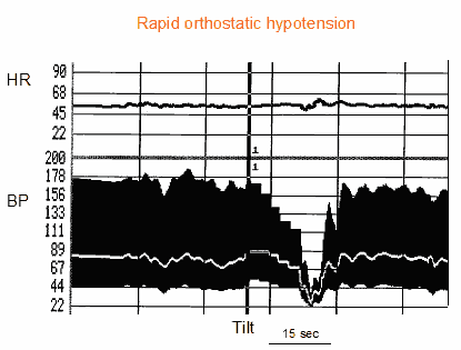
Classically, studies dealing with orthostatic intolerance have focused on patients with Rapid orthostatic hypotension due to autonomic failure. The definition commonly used is of a systolic blood pressure fall >20 mmHg or to below 90 mmHg within 3 minutes, with symptoms of cerebral hypoperfusion (5). This definition comes from a consensus designed to be used in patients with autonomic failure. Orthostatic hypotension is more frequent in the morning. Any factors which increase venous pooling in the lower limb, such as, for example, hot weather or alcohol ingestion, also worsen orthostatic hypotension. Some patients develop a syndrome of hypertension while supine alternating with hypotension while standing, probably due to impaired vasidilatation in the supine position. The presence of supine hypertension and standing hypotension renders the treatment particularly challenging. With the progression of the disease, the patient can develop chronotropic insufficiency which determines a relatively constant heart rate during the change of position or during effort (Figure 1).
Figure 1. Tilt testing in a patient with rapid orthostatic hypotension due to primary autonomic failure. From top to bottom are shown heart rate curve and blood pressure curves (systolic, diastolic and mean). There is an absence of adaptation of blood pressure to the upright position. Blood pressure declines slightly and progressively throughout the test and the patient has severe presyncopal symptoms. Heart rate is unchanged suggesting failure of cardiac adaptation to the upright position (chronotropic insufficiency)
Initial orthostatic hypotension is defined as a transient systolic blood pressure decrease within 15 s after standing >40 mmHg and disappearance within 20-30 s. There is an immediate increase in heart rate (6). Everything else is normal. Another consequence are the symptoms of a brief feeling of light-headedness accompanied sometimes by visual complaints almost immediately after standing-up for a very short duration. It is caused by a temporal mismatch between cardiac output and vascular resistance. There are some groups that are especially at risk for initial orthostatic hypotension. The first group includes young patients with an asthenic habitus. Often those patients also have postural orthostatic tachycardia and a tendency to faint during prolonged standing. Whether additional mechanical factors, such as compression of the vena cava while standing up play a role is unknown. A second group of patients includes those taking medications interfering with vasoconstrictor mechanisms, such as beta-blockers or sympathetic outflow blocking agents and psychiatric medication (6).
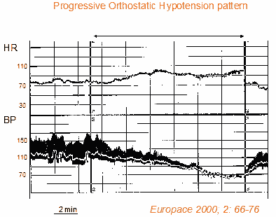
Progressive orthostatic hypotension is a condition different from classical rapid autonomic failure. Progressive orthostatic hypotension is characterised by a slow progressive decrease in systolic blood pressure (together with compensatory heart rate increases) upon the assumption of a standing position. Typically these patients remain asymptomatic initially after standing and develop hypotensive symptoms which cause orthostatic intolerance after a few minutes of standing. Progressive orthostatic hypotension also differs from neurally-mediated reflex syncopes because of the absence of vagal bradycardia which usually characterises the vasovagal reflex. Progressive orthostatic hypotension is commonly seen in the elderly because of age-related impairment in baroreflex mediated vasoconstriction and chronotropic responses of the heart, as well as to the deterioration of the diastolic filling of the heart. These patients usually have one or more associated comorbid conditions and are under treatment with vasoactive drug/s (7,8). This form of orthostatic hypotension is frequently diagnosed by tilt testing which shows the typical patterns of decreases in systolic blood pressure over several minutes (together with compensatory heart rate increases) (Figure 2).
Figure 2. Tilt testing in a patient with progressive orthostatic hypotension. From top to botton heart rate curve and blood pressure curves (systolic, diastolic and mean) are shown. There is an absence of adaptation of blood pressure to the upright position. Blood pressure declines slightly and progressively throughout the test and the patient has severe presyncopal symptoms. Heart rate continuously rises until the end of the test. There is no clear vasovagal reaction.
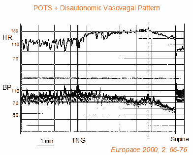
Postural orthostatic tachycardia syndrome mainly affects females (5:1 ratio with males) aged 15-50 years. This form differs from progressive orthostatic hypotension because the increase in heart rate is greater and there is less decrease in blood pressure (Figure 3). Therefore, the most frequent symptoms are palpitations and weakness. Modest physical activity can amplify the symptomatology greatly and even limit the most common daily activities. Sometimes there is hyperventilation, anxiety, loss of concentration and memory, tremors, disturbances of gastric motility and headaches, which may mistakenly suggest a diagnosis of panic disorder and chronic anxious state rather than that of postural orthostatic tachycardia syndrome. Since there isn’t any severe autonomic failure, physical examination is inconclusive and the patient is told that “everything is normal” (2,3).
Figure 3. Tilt testing in a patient with postural orthostatic intolerance syndrome (POTS). From top to botton are shown heart rate curves and blood pressure curves (systolic, diastolic and mean). During the passive phase, the heart rate increases up to 135 bpm and there is a slight decrease in blood pressure which indicates an incomplete cardiovascular adaptation to the upright position; the heart rate increase partially compensates the reduction in blood flow caused by hypotension and contributes to maintaning a cerebral flow sufficient to avoid syncope. In this phase, the patient has symptoms due to mild cerebral hypoperfusion (vertigo, weakness) and tachycardia (palpitations). The administration of 0.4 mg of nitroglycerin (TNT) causes a further increase in heart rate to 160 bpm and a decrease in blood pressure to 90 mmHg. These hemodynamic changes are probably the trigger of the following vasovagal reaction which is characterised by a decrease in heart rate (vagal outflow) and a sudden change in the slope of the blood pressure curve (sympathetic withdrawal).
Management
Some causes (e.g., volume depletion, drug induced) are transient problems that respond to treatment and do not have long-term consequences. Other diseases causing primary and secondary autonomic failure have long-term consequences and may potentially increase mortality - depending on the severity of the underlying disease.
In elderly patients with orthostatic hypotension, the prognosis is largely determined by co-morbid illnesses (1). It should be kept in mind that in dysautonomic disorders (as opposed to reflex syncopes), hypotensive syncope is but one aspect of a broader constellation of symptoms relating to autonomic failure. The physician should, therefore, not give the patient unrealistic expectations as to which symptoms can and cannot be eliminated. Both physician and patient should be aware that these disorders can be progressive in nature and that therapies may have to be altered over time.
It is reasonable for all patients to receive advice and education on factors that influence systemic blood pressure, such as avoiding sudden head-up postural changes, standing still for a prolonged period of time, high environmental temperature (including hot baths, showers), severe exertion, large meals, alcohol and drugs with vasodepressor properties. Ambulatory blood pressure recordings may be helpful in identifying circumstances (e.g., time of the day) when blood pressure fluctuation is most severe. These recordings may also help identify supine / nocturnal hypertension in treated patients.
Additional treatment principles used alone or in combination, are appropriate for consideration on an individual patient basis:
- Chronic expansion of intravascular volume by encouraging a higher than normal salt intake and fluid intake of 2-2.5 liters per day. Additional options include use of fludrocortisone in low dose (0.1 to 0.2 mg per day), and raising the head of the bed on blocks to permit gravitational exposure during sleep.
- Additional benefit may then be achieved with agents that increase peripheral resistance and reduce the tendency for gravitational downward displacement of central volume. A controlled study has shown that midodrine is superior to placebo (9)
- Interestingly, a number of patients with autonomic failure will be anaemic. A study (10) demonstrated that subcutaneous injections of erythropoietin while raising blood count will also produce dramatic increases in blood pressure.
- A recent clinical trial has shown that isometric counterpressure manoeuvres of the legs (leg crossing), or of the arms (hand grip and arm tensing), are able to induce a significant blood pressure increase during the phase of impending symptoms, which allows the patient to avoid or delay losing consciousness in most cases (11)
- The rationale for the use of elastic compression bandage is to apply an external counterpressure to the capacitance beds of the abdomen and legs in order to improve the venous return to the heart. Lower limb compression bandaging of 30-40 mmHg is effective in avoiding orthostatic systolic blood fall and reducing symptoms in elderly patients affected by progressive orthostatic hypotension. Home treatment based on self administered elastic leg stockings seems feasible, safe and well accepted by most patients (7).
The content of this article reflects the personal opinion of the author/s and is not necessarily the official position of the European Society of Cardiology.