In order to bring you the best possible user experience, this site uses Javascript. If you are seeing this message, it is likely that the Javascript option in your browser is disabled. For optimal viewing of this site, please ensure that Javascript is enabled for your browser.
Did you know that your browser is out of date? To get the best experience using our website we recommend that you upgrade to a newer version. Learn more.

Functional mitral regurgitation after NSTE-ACS

An article from the e-journal of the ESC Council for Cardiology Practice

The development of mitral regurgitation (MR) after a Q-wave acute myocardial infarction (AMI) is a recognised and frequent complication and its negative impact on survival has been well studied. Recent studies have demonstrated that MR is frequent after a NSTSEACS and that its presence and severity confers a worse long-term prognosis to patients. Thus, the presence of MR should be specifically assessed in every patient after both, a Q-wave AMI and a NSTSECAS.

Acute Coronary Syndromes


Mitral regurgitation (MR) is a frequent complication during and after the acute phase of a Q-wave acute myocardial infarction (AMI) and it is a predictor of long-term cardiovascular mortality (1-6). Very recent studies have demonstrated its prognostic implication after a non-Q wave AMI (7), the role of the existence of MR previous to an AMI (8) and its prognostic importance after a non-ST segment elevation acute coronary syndrome (NSTSEACS) (9).

Functional MR occurs with a structurally normal valve as a result of an altered force balance on the mitral leaflets (10). Its causal mechanisms have been studied in the acute and in the chronic stage after a Q-wave AMI. They include ischemia or scar at the level of the papillary muscles, annulus dilatation, change in the ventricular geometry causing tethering of the mitral leaflets and systolic dysfunction (5-10). Nevertheless, factors involved in the development of MR after NSTSEACS are still not well understood.

In a recent study, three hundred consecutive patients who were admitted to the Coronary Care Unit for a first NSTSEACS were evaluated and followed-up. Every patient had an established diagnosis of NSTSEACS based on the European Society of Cardiology criteria (11). All patients underwent a complete echocardiographic study before hospital discharge that included a specific evaluation of the mitral valve anatomy and function. The presence and degree of MR was evaluated using PISA method and a validated nomogram for semiquantitative estimation (12,13).

The main results of this study showed that a higher age and a lower ejection fraction are independent markers of MR development in this clinical scenario, probably due to the left ventricular remodelling that they cause. Patients were followed-up for a median time of 431 days. Survival curves showed that long-term outcome depends on MR presence and severity (figure). Furthermore, MR was found as a long-term independent predictor of bad outcome.

In this work, patients with a NSTSEACS and functional MR have larger left ventricular systolic diameters, as well as lower left ventricular ejection fractions. Hibernated or stunned myocardium might produce left ventricular remodelling in hearts with no transmural necrosis and the progressive change in LV size and shape might lead to the development of MR (7). It has been described that left ventricular dysfunction resulting from viable hibernating but recoverable myocardium has a better prognosis when revascularisation is performed and that an improvement of myocardial performance and reversal of dilatation decreases MR after surgical revascularisation (14,15). In this study (9), the development of MR was associated with a worse long-term outcome but it is not associated to a worse in-hospital outcome. This finding could be due to the fact that the deleterious action of post-NSTSEACS MR is based on a progressive left ventricular remodelling that develops over time. The results of the present study contrast with the results of a previous work (7) in which the presence of MR did not add any independent prognostic significance in the setting of non-Q wave AMI. Nevertheless, one must note that the population in that study was significantly smaller and the definition used for inclusion was the old definition of non-Q wave acute myocardial infarction instead the new definition of NSTSEACS.

Nevertheless, this study has some limitations. Pre-AMI MR is a recently described prognostic factor (8). It is not possible to make sure that MR was not present before the index NSTSEACS but it is very difficult to distinguish between pre-existing MR and new-onset MR. Furthermore, the morphology of the mitral valve was not specifically assessed or quantified in this work. Thus, the diagnosis of functional MR was performed excluding those patients with structural alterations at the level of the mitral leaflets or mitral subvalvular apparatus, following the methods used in previous reference papers (6).

CONCLUSIONS

Summarising, we can say that functional MR is a frequent complication after both, a Q-wave AMI and after a NSTSEACS. Furthermore, after any type of acute coronary syndrome, the presence and degree of MR adds long-term prognostic significance to other known negative factors. Thus, the presence of MR should be specifically assessed in every patient after a Q-wave AMI and also after a NSTSECAS. Nevertheless, until now, no therapeutic management has demonstrated a better outcome in this type of patients. Thus, further research is needed in this field.


Figure: Long-term outcome survival curves.
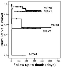
Figure legends :
MR: mitral regurgitation. 0: absence of MR or trace MR; 1: mild MR; 2= mild to moderate MR; 3: moderate MR; 4: severe MR. (adapted from Pérez de Isla L et al. Prognostic significance of functional mitral regurgitation after a first non-ST-segment elevation acute coronary síndrome. Eur Heart J 2006 27: 2655-2660).

The content of this article reflects the personal opinion of the author/s and is not necessarily the official position of the European Society of Cardiology.

References


1.- Tcheng JE, Jackman JD, Nelson CL, Gardner LH, Smith LR, Rankin JS, Califf RM, Stack RS. Outcome of  patients sustaining acute ischemic mitral regurgitation during myocardial infarction. Ann Intern Med 1992;117:18- 24.

2.- Lehmann KG, Francis CK, Dodge HT, and the TIMI Study Group. Mitral regurgitation in the early myocardial infarction. Ann Intern Med 1992;117:10- 17.


3.- Lamas GA, Mitchell GF, Flaker GC, Smith SC, Gersc BJ, Basta L, Moyé L, Braunwald E, Pfeffer MA. Clinical significance of mitral regurgitation after acute myocardial infarction. Circulation 1997;96:827- 833.


4.-Ma HH, Honma H, Munakata K, Hayakawa H. Mitral insufficiency as a complication of acute myocardial infarction and left ventricular remodeling. Jpn Circ J 1997;61:912- 920.


5.- Feinberg MS, Schwammenthal E, Shlizerman L, Porter A, Hod H, Freimark D, Matezky S, Boyko V, Mandelzweig L, Vered Z, Behar S, Sagie A. Prognostic significance of mild mitral regurgitation by color Doppler echocardiography in acute myocardial infarction. Am J Cardiol 2000;86:903- 907.


6.- Grigioni F, Enriquez-Sarano M, Zehr KJ, Bailey KR, Tajik AJ. Ischemic mitral regurgitation. Long-term outcome and prognostic implications with quantitative Doppler assesment. Circulation 2001;103:1759- 1764.


7.- Perez de Isla L, Zamorano J, Martinez Quesada M, Corros C, Ortiz P, Almeria C, Rodrigo JL, Aubele AL, Fernandez-Ortiz A, Macaya C. Prognostic significance of ischemic mitral regurgitation after non-Q-wave acute myocardial infarction. J Heart Valve Dis. 2005 Nov;14(6):742-8.


8.- Zamorano J, Pérez de Isla L, Oliveros L, Almería C, Rodrigo JL, Aubele A, Banchs J, Macaya C. Prognostic Influence of Mitral Regurgitation Prior to a First Myocardial Infarction. Eur Heart J. 2005 Feb;26(4):343-9.


9.- Pérez de Isla L, Zamorano J, Quezada M, Almería C, Rodrigo JL, Serra V, Garcia Rubira JC, Fernandez Ortiz A, Macaya C. Prognostic significance of functional mitral regurgitation after a first non-ST-segment elevation acute coronary syndrome. Eur Heart J 2006 27: 2655-2660.


10.- He S, Fontaine AA, Schwammenthal E, Yoganathan AP, Levine RA. Integrated mechanism for functional mitral regurgitation. Leaflet restriction versus coapting force: in vitro studies. Circulation 1997;96:1826- 1834.


11.- Bertrand ME, Simoons ML, Fox KA, Wallentin LC, Hamm CW, McFadden E, De Feyter PJ, Specchia G, Ruzyllo W. Management of acute coronary syndromes in patients presenting without persistent ST-segment elevation. The Task Force on the Management of Acute Coronary Syndromes of the European Society of Cardiology. European Heart Journal (2002) 23, 1809–1840.


12.- Enriquez-Sarano M, Miller FA Jr, Hayes SN, Bailey KR, Tajik AJ, Seward JB. Effective mitral regurgitant orifice area: clinical use and pitfalls of the proximal isovelocitysurface area method. J Am Coll Cardiol 1995;25:703- 709.


13.- Moya JL, Catalan MP, Garcia-Lledo A, Pey J, Barcia F, Asin E. A semiquantitative method based on proximal convergence zone to estimate the severity of the mitral regurgitation: design and clinical application. Eur J Echocardiogr. 2001 Sep;2(3):163-9.

14.- Pu M, Thomas JD, Gillinov MA, Griffin BP, Brunken RC. Importance of ischemic and viable myocardium for patients with chronic ischemic mitral regurgitation and left ventricular dysfunction. Am J Cardiol 2003;92:862- 864.


15.- Campwala SZ, Wang N, Pai RG. Factors affecting regression of ischemic mitral regurgitation following isolated coronary artery bypass surgery. J Am Coll Cardiol 2003; supplA:504A.


VolumeNumber:

Vol6 N°04

Notes to editor


*Dr Leopoldo Perez de Isla and Pr Jose Zamorano.
Instituto Cardiovascular. Hospital Clínico San Carlos. Madrid. Spain.
*Member of the European Association of Echocardiography (EAE), and President-Elect

Correspondence:
Leopoldo Perez de Isla
Echocardiographic Laboratory
Hospital Clínico San Carlos
Plaza Cristo Rey
28040-Madrid, Spain
Tel: 0034913303290
Fax: 0034913303290
leopisla@hotmail.com

The content of this article reflects the personal opinion of the author/s and is not necessarily the official position of the European Society of Cardiology.